Amniocentesis: what is and how is this diagnostic test performed?
Pregnancy and pregnancy are very delicate stages, because in this biological process the new organism begins to develop. That is why, from a medical point of view, it is important know as much as possible about what is happening in the development of the fetus , to be able to intervene as soon as possible in the case of congenital diseases.
Amniocentesis is the procedure that doctors perform to obtain this early information and be able to make an early diagnosis during pregnancy. Throughout this article we will review everything that is necessary to know about this test: what amniocentesis is, what its functions are, how it is performed and what are the risks to be taken into account.
- Related article: "The 3 phases of intrauterine or prenatal development: from the zygote to the fetus
What is an amniocentesis?
We call amniocentesis a a type of prenatal test in which an early diagnosis is made through a medical procedure of chromosomal diseases and fetal infections and that secondarily, also helps us to know what is the sex of the baby before birth.
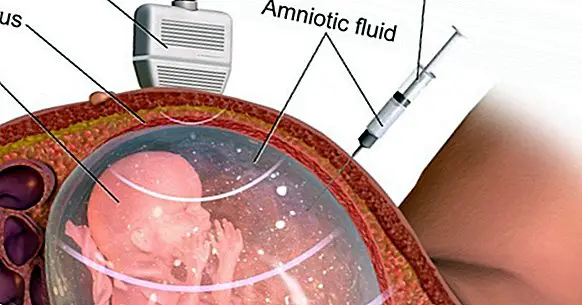
To understand how it works, you must first know that throughout the gestation stage the fetus is surrounded by a substance called amniotic fluid , whose composition has fetal cells. From the observation of this fact, the scientific community applied to the clinical field has discovered that the amniotic fluid is able to give us useful information about the health of the baby months before the birth occurs. Amniocentesis focuses on the analysis of that substance and its components.
At the time of performing the amniocentesis a small sample of amniotic fluid is obtained through the use of a needle that is inserted into the woman's abdomen at the same time that an ultrasound is being performed with which you can monitor the process. Secondly, the sample of amniotic fluid obtained is analyzed in the laboratory, a context in which the DNA of the fetus is studied to see if there are genetic anomalies in it.
In which cases is it done?
This prenatal test is only offered to women who present a significant risk of genetic disease. In most cases, the main reason to perform an amniocentesis is to know if the fetus has any chromosomal or genetic abnormality as it may occur in Down syndrome. As a general rule, this diagnostic procedure is scheduled between weeks 15 and 18 of pregnancy .
Therefore, it is not always necessary to do it, in most cases it is only done in pregnant women in which the baby presents some risk of developing a genetic pathology. The reason why not all women are treated is that they are a fairly invasive test that carries a small risk of spontaneous abortion .
Given that amniocentesis is associated with certain risks, before performing it, a complete anatomic ultrasound is performed to detect anomalies in the baby. In cases where there are reasons to suspect the existence of genetic or chromosomal alterations , the amniocentesis will be performed.
Functions of this test: what is it for?
The main cases in which an amniocentesis is required include:
- A family history of congenital defects .
- Abnormal results in ultrasound tests.
- Women with pregnancies or children where there alterations of birth or pregnancy .
Unfortunately, amniocentesis does not detect all possible birth defects. However, the ultrasound test performed at the same time, can detect congenital defects that can not be reported in amniocentesis such as cleft lip, heart defects, cleft palate or clubfoot.
However, the risk of some birth defects that are not detected through either of the two diagnostic tests can not be ruled out. In general, the main diseases detected by amniocentesis are:
- Muscular dystrophy.
- Cystic fibrosis.
- Sickle cell disease .
- Down's Syndrome.
- Alterations in the neural tube , as occurs in spina bifida.
- Tay-Sachs disease and related.
Finally, the accuracy of amniocentesis is approximately 99.4%, so although it has certain dangers it is very useful in cases in which there is a real suspicion of a fetal anomaly.
How do doctors do it?
After having cleaned with an antiseptic the area of the abdomen where the needle will be inserted and administering a local anesthetic to relieve the pain of the puncture, the medical team locates the position of the fetus and the placenta using an ultrasound. Turning around these images, a very fine needle is inserted through the abdominal wall of the mother , the wall of the uterus and the amniotic sac, trying to get the tip away from the fetus.
Then a small amount of liquid is extracted, of plus or minus 20 ml, and this sample is sent to the laboratory in which the analysis will be carried out. In this space, the fetal cells are separated from the rest of the elements present in the amniotic fluid.
These cells are cultured, fixed and stained so that they can be observed correctly through the microscope. A) Yes, the chromosomes are examined anomalies .
As for the baby and its environment, the puncture seals and the fluid in the amniotic sac regenerate during the following 24-48 hours. The mother should go home and rest for the rest of the day, avoiding physical exercise. In a matter of one day, you can return to normal life unless the doctor indicates otherwise.
The risks
Despite the fact that safety measures in medicine have advanced a great deal in this area as well, Amniocentesis always presents risks . The risk of spontaneous abortion is the most notorious, although it occurs only in 1% of cases.
The possibility of premature birth, injuries and malformations in the fetus is also an aspect to be taken into account.
Bibliographic references:
- Carlson, L. M. & Vora, N. L. (2017). Prenatal Diagnosis: Screening and Diagnostic Tools. Obstetrics and Gynecology Clinics of North America, 44 (2): 245-256.
- Seeds, J. W. (2004). Diagnostic mid trimester amniocentesis: how safe ?. American Journal of Obstetrics and Gynecology, 191 (2): 607-15.
- Underwood, M. A, Gilbert, W. M, Sherman, M. P. (2005). "Amniotic Fluid: Not Just Fetal Urine Anymore". Journal of Perinatology. 25 (5): pp. 341-348.