Cerebral hemangioma: causes, symptoms and treatment
Our vascular system is a fundamental element for our survival, since it allows the oxygen and nutrients our cells need to reach them through the blood. Thus, our life can be in serious danger if this system becomes damaged, depending on the area and the type of blood vessels affected.
Sometimes malformations or neoplasms also occur in the form of uncontrolled and disorganized growths of blood vessels which can also pose a danger especially if they occur in areas such as the brain. It's what happens with the brain hemangioma .
- Related article: "The differences between syndrome, disorder and disease"
What is a hemangioma?
A hemangioma is a type of neoplasia or uncontrolled growth of blood vessel cells . They could be considered a type of benign tumor of the vascular system, which, like other tumors, can grow even though they do not have malignancy.
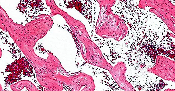
The hemangioma itself can appear in different regions of the body, such as the skin, but also in areas such as the lung, stomach or brain. They can appear as nodules or endothelial cavities filled with blood, which can burst and produce effusions with great ease.
While in some cases they may not cause complications when they occur in organs such as the skin, when they appear in other organs such as lung or brain They can have devastating consequences.
- Related article: "The 16 most common mental disorders"
The cerebral hemangioma
The cerebral hemangioma, also called cavernous angioma, is a type of hemangioma that arises in some areas of the brain. They are generally considered to be a product of congenital malformations that are suffered from childhood and during development. In the case of cerebral hemangioma, the consequences of this one producing a bleed can be really dangerous and even the death of the subject.
This is so because, similar to the aneurysm, the presence of a hemorrhage within the brain can drown and drown nearby nerve cells , causing his death and loss of functions. And even if the bleeding is self-contained within the nodule itself, it can cause it to grow and compress areas of the brain. It can also cause a stroke.
Depending on the location, the consequences may be one or the other. Headache, tiredness, seizures, sensory problems are common. The presence of nausea and vomiting is also frequent. If they occur in the brainstem, they can affect cardiorespiratory, digestive function or even the death of the patient.
In most cases they tend to appear supratentorially (that is, above the cerebellum) in the frontal or temporal lobes, although they can also arise in the cerebellum and in the pons. Movement, language and the ability to reason could be impaired. In some cases, however, the cerebral hemangioma remains asymptomatic, although there is a risk of bleeding.
Causes
The cerebral hemangioma it is usually a congenital malformation in the form of neoplasia . Its causes are currently little known. However, it has been detected that there are variations such as familial cavernous angioma in which the problem has been associated with genetic mutations on chromosome 7. In other cases in which it appears sporadically, it can be due to de novo genetic mutations.
Hemangioma treatment
Treating the presence of a brain hemangioma can be complex, and you have to take into account the possibility of complications.
In cases in which the hemangioma remains stable and does not cause problems or bleeding, treatment may not be carried out beyond the performance of a periodic control of the case.
Otherwise, the main objective of the interventions in this type of malformations is that of make it stop circulating the blood for them , so that the risks of bleeding are avoided and can be eliminated.
Since the surgery itself can be dangerous, it is usually reserved for cases in which bleeding is occurring and the possible benefits outweigh the risks. The resection of the malformation must be complete, or else there is a risk of increasing the bleeding.
For this, several techniques can be used, the embolization of the hemangioma is frequent . This procedure is based on the application of substances that clog the blood vessels, so that the blood vessel stops carrying blood and becomes encyst. Once frozen, the nodules are removed. They can also be treated with corticosteroids if they are in a slow growth phase, to reduce their size by reducing the level of inflammation of the angioma.
Bibliographic references:
- Cortés, J.J .; Bernabé, J.M .; Riera, N. and Arenas, J.J. (2009). Intracranial cavernous angiomas. Radiology; 51: 190-193. Alicante, Spain
- Isla, A .; Alvarez, F .; Muñoz, J .; Nos, J. and García-Blázquez, M. (1995). Treatment of cavernous angiomas. Neurosurgery; 6 (2): 138-145. Hospital La Paz. Madrid.
- Fritschi, J.A .; Reulen, H.J .; Spetzler, R.F. & Zabramski, J.M. (1994). Cavernous malformations of the brain stem. A review of 139 cases. Acta Neurochir (Wien). 1994; 130 (1-4): 35-46. Review