Cranial nerves: the 12 nerves that leave the brain
In general, it can be said that the human brain communicates with almost all the nerves of the brain through the spinal cord.
Thus, for example, the information that reaches us about what we touch with our hands is picked up by nerves that run along the arm until we reach the spinal cord, and from there to the brain, from where the order to continue examining the object will be issued. This efferent order will also leave the brain through the spinal cord, and it will reach the corresponding arm through the nerve fibers that come out of it.
However, this is not a rule that is always fulfilled, since there are also some nerves that come directly from the brain, without being born in the spinal cord. It's about cranial nerves, or cranial nerves , which arise from the lower part of the brain and reach their target areas through small holes distributed at the base of the skull. From these orifices, the cranial pairs communicate with peripheral areas.
In addition, although it may seem strange, not all these cranial nerves have the function of reaching areas and organs that are in the head. Some extend to the neck and even the abdomen area.
How are the cranial pairs classified and distributed?
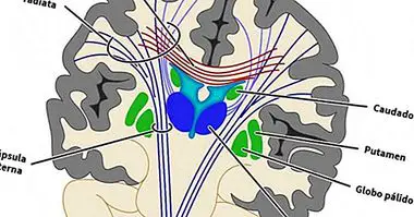
The cranial nerves they are so called because they are counted in pairs, as there is one on both the right and left sides of the brain . Thus, there are twelve cranial nerves pointing to the right hemisphere and another twelve pointing to the left, symmetrically.
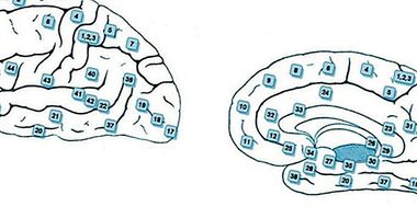
Each pair is numbered with a Roman numeral according to whether the position from which they leave the brain more or less near the frontal zone. In fact, the cranial nerves can be grouped and classified into categories according to two criteria : the place from which they start and their function.
Cranial pairs classified according to their position
- Starting from areas that are above the brainstem are the pairs I and II .
- Starting from the mesencephalon (the upper part of the brainstem), there are cranial pairs III and IV .
- Starting from the Varolio bridge (or trunk-bridge), there are cranial nerves V, VI, VII and VIII .
- Starting from the medulla oblongata (in the lower part of the brainstem) are the nerves IX, X, XI and XII .
Cranial cranial classified according to their function
- Sensitive : pairs I, II and VIII.
- Related to the movements of the eyes (and its parts) and the eyelids: cranial nerves III, IV and VI.
- Related to the activation of muscles of the neck and tongue : the cranial pairs XI and XII.
- Mixed cranial nerves : the pairs V, VII, IX and X.
- Parasympathetic fibers : nerves III, VII, IX and X.

What are the cranial nerves?
We will now know what are the cranial pairs one by one, and their main functions.
1. Olfactory nerve (cranial nerve I)
As its name suggests, this cranial nerve is dedicated to transmit specifically nerve information about what is detected through the sense of smell , and therefore it is an afferent fiber. It is the shortest of the cranial pairs, since its place of destination is very close to the area of the brain from which it arises.
2. Optic nerve (cranial pair II)
It is also part of the afferent fibers, and is responsible for transmitting to the brain the visual information that is collected from the eye . It arises from the diencephalon.
3. Oculomotor nerve (cranial nerve III)
Also know as common ocular motor nerve, this cranial nerve sends orders to the majority of muscles that are involved in the movement of the eyes , and causes the pupil to dilate or contract.
4. Trochlear nerve, or pathetic nerve (cranial nerve IV)
Like the oculomotor nerve, this cranial pair deals with the movement of the eyes . In particular, it sends signals to the superior oblique muscle of the eye. The place from which this pair of nerves arises is the mesencephalon.
5. Trigeminal nerve (cranial nerve V)
It is one of the mixed cranial nerves, because It has both motor and sensory functions . In its facet of motor nerve, it sends orders to muscles in charge of performing the movements of mastication, while as sensory cranial nerve it gathers tactile, proprioceptive and pain information from several areas of the face and mouth.
6. Abducent nerve (cranial nerve VI)
This is another of the cranial nerves responsible for making the eye move . In particular, it is responsible for producing abduction, that is, the eye moves to the opposite side to where the nose is.
7. Facial nerve (cranial nerve VII)
It is one of the mixed cranial nerves. It is responsible for sending orders to facial muscles dedicated to creating facial expressions (allowing thus to socialize and communicate correctly) as to the lacrimal and salivary glands. It also collects taste data from the language.
8. Vestibulocochlear nerve (cranial nerve VIII)
It is one of the sensory cranial nerves, and collects information from the auditory zone . Specifically, it receives data relative to what is heard and to the position in which we are in relation to the center of gravity, which allows us to maintain balance.
9. Glossopharyngeal nerve (cranial nerve IV)
It is a sensitive and motor nerve and, as its name suggests, it has influence on both the tongue and the pharynx (the tube that connects the mouth to the stomach). It receives information from the taste buds of the tongue, but also sends orders to both the parotid gland (salivary gland) and neck muscles that facilitate the swallowing action.
10. Vagus nerve (cranial pair X)
This cranial pair takes orders to most of the pharyngeal and laryngeal muscles , sends nerve fibers from the sympathetic system to viscera that are in the area of our abdomen and receives taste information that comes from the epiglottis. Like the glossopharyngeal nerve, it intervenes in the action of swallowing, so it has a lot of relevance given the importance of this vital function.
11. Accessory nerve (cranial nerve XI)
To this cranial pair also it is known as a spinal nerve .
It is one of the pure cranial nerves, and activates the trapezius and sternocleidomastoid muscles , that intervene in the movement of the head and shoulders, so that their signals are noticed in part of the upper part of the thorax. Specifically, it allows the head to be decanted to one side and to be able to lean backwards.
12. Hypoglossal nerve (cranial nerve XII)
Like the vagus nerve and the glossopharyngeal nerve, to ctiva muscles of the tongue and participates in the action of swallowing . Thus, it works together with cranial nerves IX and X to allow swallowing to be performed correctly, which is fundamental for the good state of the organism.