Endoderm: parts and development in pregnancy
The development and growth of the human body is an extremely complex and fascinating process in which the different structures work with millimetric precision to give rise to the birth of different organs and body systems.
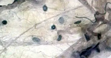
One of these structures is the endoderm , a layer or stratum of tissue of which we will discuss throughout this article. This layer is one of the oldest biological parts at the developmental level and gives rise to important vital organs such as those found in the digestive system.
- You may be interested: "Neurulation: the neural tube formation process"
What is the endoderm?
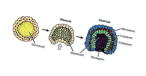
The endoderm refers to layer of innermost tissue of the three layers that develop during embryonic growth of the animals. These strata known as germinative layers are the ectoderm, which is the outermost layer and the mesoderm or middle layer.
However, it is necessary to specify that not all species have these three germinative layers. According to each animal group, the embryonic cells can derive in two or three layers, forming diblastic and triblastic beings respectively. Even so, they all have the endoderm layer, which is below the rest.
In the case of the endoderm, it appears around the third week of gestation, being considered as one of the oldest layers within the process of embryonic differentiation. Further, it is from this stratum of cells that many vital organs are born for the survival of the adult living being.
From this stratum germinate, the great part of the most important internal organs will be formed. Some of them are the alveoli found in the lungs, the entire digestive system as well as their secretory glands, the epithelia of some glands such as the thyroid or thymus, and finally some parts of the kidneys, bladder and urethra.
- Related article: "How to take care during the first month of pregnancy: 9 tips"
How is it developed?
During the early stages of embryonic development, the embryo is formed by a single layer of cells. Then, it is folded on itself in a process called gastrulation, thanks to which the first cellular layers are born. The first of these layers to appear is that of the endoderm.
Around the second week of gestation, a group of migrating cellular organisms slide to the hypoblast cells , an internal mass formed by cubic cells, and becomes the final endodermal layer.
The next phase in the evolution of the embryo is called organogenesis. This is responsible for producing the corresponding changes in the embryonic layers and give way to the formation of suitable organs and tissues.
As indicated above, in the case of the endoderm, this will lead to different organs of the digestive and respiratory system , as well as the epithelial envelope of some parts of the body. However, it is necessary to specify that these bodies are not about the definitive structures but about the primitive members that are still to be fully developed.
Types of endoderm
Following the differentiation of the embryonic body, the endoderm is divided into two parts that have their own characteristics. These parts are the embryonic endoderm and the extraembryonic endoderm. These two divisions are connected by a wide orifice that, later, it will become the umbilical cord .
1. Embryonic endoderm
The embryonic endoderm is the section of the endodermal layer that will give rise to the internal structures of the embryo, forming the primary intestine. In addition, this embryonic stratum works together with the mesodermal layer to form the notochord . When this structure is fully developed it is the main one in charge of emitting the necessary signals to enable migration and cell differentiation; an extremely important process to enable the formation of organic structures such as the brain.
From here, the notochord and the endoderm perform a parallel development in which the first generates a series of folds that will form the cranial, caudal and lateral axes of the embryo; while the folds of the endoderm are staying inside the organism forming the intestinal tube.
2. Extraembryonic endoderm
The second division of the endoderm is that which remains outside the embryoOrmando the well-known yolk sac . This membranous annex is connected to the embryo, supplying sufficient nutrients and oxygen, as well as discarding metabolic waste.
However, this division of embryonic endoderm does not remain until the end of embryonic development but usually disappears around the tenth week of gestation.
Sections of the intestinal tube
In the previous section it was mentioned that the embryonic endoderm gives rise to a structure called intestinal tube. This structure can be differentiated in turn into different sections that may correspond to both the embryonic endoderm and the extraembryonic endoderm. These sections are:
1. Cranial bowel
Known as cranial or internal bowel , this structure is located inside the skull of the embryo. During the early stages of development this forms the oropharyngeal membrane, which gradually transforms into the pharynx. Next, the lower limit forms a structure known as the respiratory tract.
Finally, the intestinal tube it dilates until it becomes what will finally correspond to the stomach .
2. caudal intestine
Located inside the caudal fold is the precursor of the allantoic membrane . An extraembryonic layer that appears by forming folds located next to the yolk sac.
3. Middle intestine
Finally, the midgut is located between the cranial and caudal structures. Its extension dilates to reach the yolk sac through the umbilical cord. Thanks to him, the embryo is supplied with sufficient nutrients from the mother's organism and allow their correct development.
In what organs is it transformed?
It has already been mentioned on multiple occasions that the endoderm is the primitive structure thanks to which arise a large part of the organs and body structures, a process known as organogenesis , that is, the birth of the organs.
This process of development of the definitive organs usually takes place between the veal and the eighth week of management. However, it has been proven that in the individuals identifiable organs can be observed from the fifth week.
To be more concrete, of the endodermal layer derive these structures :
- Yolk sac.
- Alantoides
- Respiratory tract , specifically the bronchi and pulmonary alveoli.
- Epithelium and connective tissue of the tonsils, pharynx, larynx and trachea. As well as the epithelium of lungs and certain parts of the gastrointestinal tract.
- Urinary bladder.
- Epithelium of the Eustachian tubes, ear cavities, thyroid and parathyroid glands, thymus, vagina and urethra.
- Glands of the digestive system , specifically of the gastrointestinal tract; as well as the liver, the gallbladder and the pancreas.