Nissl bodies: anatomy, characteristics and functions
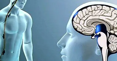
Research and exploration of the human brain and the structures that are part of it have been constant since antiquity. The neuron as a basic unit of the nervous system has been specially researched, using strategies such as the use of different stains to observe its structure.
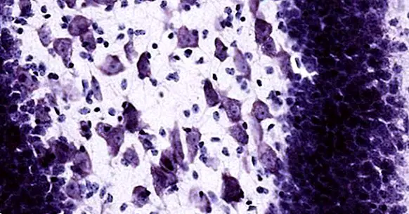
The German neurologist Franz Nissl elaborated a staining based on dyes such as toluidine blue or cresyl violet, and before his application he could observe how this substance clearly showed the existence of different structures in the neuronal cytoplasm. They had discovered what we now know as corpuscles or bodies of Nissl .
Nissl's bodies: what are they?
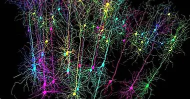
The bodies of Nissl or ergastoplasma are small structures in the form of corpuscles or granules present in neurons of the nervous system. These structures are located in the cytoplasm of the cell, and are located in specific parts of the neuron. They can be found especially in the soma or nucleus of the neuron and also in the dendrites, not found in the neuronal axon.
Nissl bodies are considered to be cumulative rough endoplasmic reticulum . In other words, they are structures formed by parallel cisterns with ribosomes (enzymatic structures made of ribosomal RNA) adhered in a spiral, in which, in addition, free polyribosomes can also be seen. These bodies only appear in eukaryotic cells, that is to say those that have a nucleus such as neurons, and have the function of secreting proteins.
They are also basophil structures, characterized by the affinity and ease of staining by dyes. In these structures there a high concentration of both ribosomal and messenger RNA , the active ribosomes being attached to the latter.
They can have different sizes and be presented in different amounts depending on the type of neuron. Those that are part of the ganglia of the autonomic nervous system tend to be small, while other large neurons tend to have larger Nissl bodies.
- Maybe you're interested: "Differences between DNA and RNA"
Function of these structures
Nissl bodies, as conglomerates of rough endoplasmic reticulum in which ribosomes are observed and in which both ribosomal and messenger RNA can be found, Their main function is the synthesis and transport of proteins inside the cell. Specifically, the part of the Nissl bodies that have the most action when generating proteins to be used inside the cell are free polyribosomes.
The proteins secreted by these bodies are fundamental in the face of transmit nerve impulses between neurons , as well as participate in the generation of neurotransmitters.
In addition, Nissl's body plays an important role in maintaining the health of the cell, by allowing the regeneration of structures damaged by the neuron's own activity or by external factors.
Chromatolysis as a defense against neuronal damage
Nissl bodies can be damaged by possible injuries or pathologies. Neural damages such as those caused by trauma and diseases they can generate damage to the axon.
The presence of damage to the axon causes the neuron to react by swelling and displacing the nucleus to move it away from the lesion. It also acts by giving a response called chromatolysis, in which the Nissl bodies move from the neuronal cytoplasm to the injured area in order to repair it. The reorganization and regeneration of the axon is allowed, so that the functionality of the neuron is recovered, but while this occurs Nissl bodies dissolve . Fortunately, if recovery of the neuron is achieved, chromatolysis ceases and the cytoplasm can recover and form new bodies.
This reaction may appear as we have said before injuries caused by trauma, but they have also been observed in various disorders. It is common to observe its appearance in neurodegenerative processes such as dementia due to Pick disease or Alzheimer's disease (in fact, the changes in the cytoplasm that cause this event is usually considered as a sign of neuronal degeneration, so that its occurrence can be a possible sign of danger), in Wernicke encephalopathy of Wernicke-Korsakoff syndrome, diseases such as porphyria or some infectious diseases. It can also be observed in normative aging or in the face of a situation of great continuing stress for the individual.
Bibliographic references:
- Gómez, M. (2012). Psychobiology CEDE Preparation Manual PIR.12. CEDE: Madrid-
- Ramón y Cajal, S. (2007). Histology of the nervous system of man and vertebrates.Volume i. Ministry of Health. Madrid.