Somatosensory cortex: parts, functions and associated pathologies
Each region of the human brain has specific functions and charges, ranging from controlling the voluntary and involuntary movement of our muscles, enabling the production of language or receiving and interpreting all the sensations that come from our environment.
This last function is possible thanks to the somatosensory cortex , a very specific brain area that we will talk about throughout this article. In addition, we will describe their specific regions and what happens when they suffer some type of injury.
- Related article: "Parts of the human brain (and functions)"
What is the somatosensory cortex?
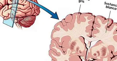
The concept of somatosensory cortex refers to a specific brain area located in the parietal lobe . As its name suggests, this lobe is under the parietal bone of the skull and in terms of size, the parietal lobe is one of the largest among those that make up the skull.
Among all the areas that define the temporal lobe, the somatosensory cortex is responsible for processing and treating sensory information that comes from the dermis, muscles and joints.
Among the functions of the somatosensory cortex, is that of receive and interpret all that information that comes from the tactile system . The sensations of pain, temperature, pressure, as well as the ability to perceive the size, texture and shape of objects are also possible thanks to this section of the cerebral cortex.
In the same way, the somatosensory area of the brain is also responsible for receiving and transmitting information related to the position in which our body is located with respect to the space that surrounds it.
In summary, the main functions of the somatosensory cortex are:
- Processing of the sensations of pain .
- Processing of tactile information.
- Processing of thermal sensations.
- Processing of vibratory sensations.
- Voluntary movements of the hands.
- Movement of the orofacial musculature.
- Voluntary swallow .
Finally, each of the specific areas within the somatosensory cortex are specialized in interpreting the sensory information of certain specific areas of our body. These areas are divided between the primary somatosensory area and the somatosensory association area, which are described in the third and fourth points of this same article.
- Maybe you're interested: "Nociceptors (pain receptors): definition and types"
Layers of the somatosensory cortex
Like the rest of the cerebral cortex, the somatosensory cortex is formed by a series of layers that have their own and well-defined functions. Specifically, The somatosensory cortex is formed by six layers of nervous tissue .
The first of these layers is the outer one, the one that is closest to the brain surface. Among its functions is to send sensitive signals that stimulate the fourth layer. In addition, both the first tissue layer and the second receive signals that control the level of excitability of the sensory system.
As for the second and third layer of the somatosensory cortex, the neurons that compose it are responsible for sending, through the corpus callosum, signals to the related regions of the corresponding cerebral cortex of the opposite hemisphere.
Finally, the fifth and the sixth layer have as main and only function send neuronal signals to the deeper areas of the nervous system .
Primary somatosensory area
As specified at the beginning of the article, the somatosensory cortex is divided into two specific areas. The first of these is the primary somatosensory area. This area is the main one in charge of the treatment of somatic sensations .
The information in which these sensations are stored is sent by the receivers that are located throughout the entire body. These receptors receive information from the outside regarding touch, pain and temperature and the information that allows us to know what position or situation our body is in. At the same moment in which these receptors perceive any of these sensations, they transmit the information to the primary somatosensory cortex through the nerve fibers found in the thalamus.
The primary somatosensory cortex refers to areas 1, 2 and 3 of the 52 brain regions described by Brodmann, which they are located concretely in the postcentral gyrus , occupying both the lateral and medial zones.
As mentioned in the first point, each of the regions of the somatosensory cortex, in this case the primary somatosensory cortex, is specialized in receiving information from a specific area of our body. This disposition is a function of the sensitivity level of the different body areas, so very sensitive areas such as the lips, hands or genitals, which have a large number of nerve endings, require many more neural circuits and an area in the cortex much more extensive.
There is a graphic or somatotopic representation of this distribution of the primary sensory cortex. This picture is known as sensory homunculus or Penfield . It depicts a map of the cerebral cortex in which they show how the different organs and senses of the body have a specific place in the brain.
Further. in the sensory homunculus the size of the organs represented is a function of the number of nerve endings that it possesses and of the functional importance of the specific area. That is, the more endings, the greater the size of the representation.
Lesions of the primary somatosensory cortex
Any type of injury or deterioration caused in this area can cause numerous alterations in the ability to perceive sensations. Among these dysfunctions are:
- Decrease or loss of pain and thermal sensations
- Alterations in the ability to perceive the position of one's own body and movements
- Impairment of sensations and tactile functions
Somatosensory association area
The second region of the somatosensory cortex is known as the somatosensory area or association cortex. It produces the union and integration of all the information corresponding to the general sensations .
Thanks to this area of the cortex, we can recognize and identify the stimuli and objects that surround us; since it allows the evaluation and understanding of the general characteristics of these.
For this, this area of association, located in areas 5 and 7 Brodmann, receives bundles of fibers that come from the primary somatosensory area and that are loaded with sensory information ready to process.
Lesions of the somatosensory cortex of association
In the cases in which this region suffers any type of damage or degradation, the ability to perceive the general sensations of the environment is maintained. However, the brain is completely incapable of integrating and give a sense to this information .
This is the case of agnosias, in which the main problem is a deficit in the recognition of objects and people.