Spinal cord: anatomy, parts and functions
When we think about the nervous system, we usually think almost exclusively about the brain.
Focusing on this organ is logical due to its special relevance, but it is often forgotten that the nervous system is precisely a system, that is, a set of interrelated elements. In other words, not everything is the brain. Moreover, within the nervous system there are two major divisions, the central nervous system and the autonomic nervous system.
Besides the king organ, in the central nervous system we can also find another great component: the spinal cord, through which most of the innervations of the body pass .
A general description: the spinal cord
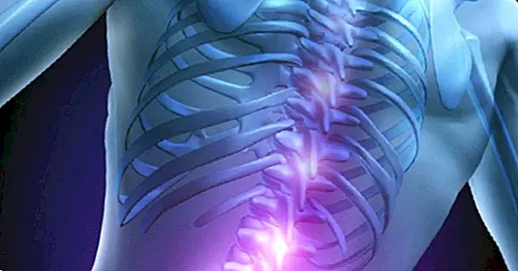
The spinal cord is the most caudal part of the central nervous system, starting in the medulla and ending in the lower back. It is the lower part of the neuroaxis, with a slightly flattened and asymmetrical cylindrical shape that, like the brain, is strongly protected by being surrounded by the spine. Likewise, it also enjoys the protection of the meninges and cerebrospinal fluid, which prevent most of the damage caused by environmental elements.
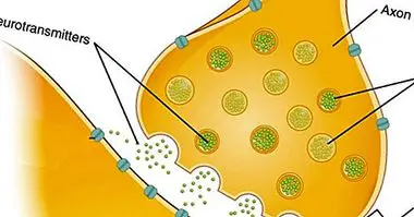
This part of the nervous system is the point of connection between the brain and the rest of the body , the vast majority of nerve fibers passing through the marrow. The transmission of information is not usually through a single neuron, but as a general rule, the neurons that make up the different nerves of the body make one or several intermediate synapses, either within the marrow itself or outside it (as with the neurons of the nerve ganglia).
The spinal cord receives both afferents and eferences , that is, it has both neurons that receive information from the recipients of the different organs and structures and others that send information and commands to those areas.
Neuroanatomical configuration
Although the division into vertebrae has more to do with the configuration of the spine, that is, the bone protection of the spine which in turn serves as a supporter of the body position, it may be useful to take it into consideration in order to locate the situation of the spine. the parts of the medulla that innervate the different body areas.
Most human beings are born with a total of 33 vertebrae , including seven cervical vertebrae, twelve thoracic vertebrae, five lumbar vertebrae, five sacral vertebrae and four coccygeal vertebrae. As we develop, the number is reduced by merging the lower ones to form the sacral and coccygeal bones, going on to consider only the first 24 vertebrae, ending in L5 or lumbar 5. The beginning of the spinal cord is located a little earlier of its covering by the vertebral column, being adhered to the medulla oblongata. The point where the marrow ends can vary from one person to another, culminating generally between vertebrae L1 and L3.
In general, the nerve connections correspond to the marrow correspond to the area where they are located. Thus, in the part of the cord located in between the thoracic vertebrae are the nerve connections that innervate the thorax, and so on. In regard to the nerves that connect with the cord, we have a total of thirty-one pairs, eight cervical, twelve thoracic, five lumbar, five sacral and one coccygeal. A point to note is the presence of two areas in which the medulla is somewhat wider, because in these areas are the nerve connections with the extremities.
Between the vertebrae C4 and T1 there is a somewhat wider area than the rest of the medulla. This area, known as cervical intumescence, is thicker because in this place are the nerve connections that connect with the upper extremities.
Towards the inferior end of the marrow a thickening can be observed, between which it goes from the vertebra T11 to the L1, denominated lumbosacral intumescence. It is the part of the medulla that innervates the lower extremities, and which together with the so-called horsetail connects with the parts of the body located at the lower end.
Regarding the aforementioned horsetail, which receives its name due to the similarity of its shape with the tail of said animal, is the set of nerve fibers that connect with the spinal nerves. This form is due to the fact that the spinal cord is shorter than the spine, so that the areas below the lumbar area must project their nerve endings to the spinal nerves located below it.

Parts of the marrow
It has been observed that the medulla has different nervous connections that innervate different areas of the body. However, it may be of interest to analyze the internal structure of the spinal cord.
As in the brain, in the marrow we find both gray substance and white matter . However, the arrangement is reversed, with the white substance located in an external position and the gray substance in the inner part of the medulla. Generally, the transmission of information occurs in an ipsilateral manner, that is, the right side of the body is treated by the left side of the spinal cord while the left side is treated with the right side.
Gray substance
The gray substance has this coloration because it is a set of somas or nuclei of neurons, which project their axons to other areas. That is to say, it is in these zones where the bodies of neurons accumulate, centers of information processing (although not being in the brain that processing is very shallow). The gray substance is structured in different horns or antlers, being the main the ventral horn, the dorsal horn and the intermediate zone. There is also the lateral horn, but only in the thoracic area and the beginning of the lumbar.
The dorsal horn is responsible for receiving information from the systems innervated by the medullar to . In other words, it is the part of the medulla that is responsible for the external or internal stimulation detected by the receptors to be sent to the brain.
The ventral horn of the cord, unlike the dorsal horn, has the main function of emitting information to the nerves, causing the body to react to external or internal stimuli. Through it the voluntary movement is exercised.
With regard to the intermediate zone, there are many interneurons, which are those whose main function is to serve as a link between two other neurons. They are connection bridges between distal zones.
Although it only appears in the thoracic area and part of the lumbar region, the lateral horn is of great importance, innervating different structures and participating in the sympathetic and parasympathetic systems of the autonomic nervous system. In this sense, it plays a fundamental role in homeostasis, the process by which the organism establishes a balance or harmony between different areas of the body so that the set of organs function in a healthy and coordinated manner.
White substance
The white substance is formed mainly by the axons of the neurons, interconnecting bone and brain . It is organized in different fibers that are named after the zones with which they connect, which can be ascending or descending. In the marrow you can find three columns, the dorsal, the lateral and the ventral.
The dorsal column is mainly formed by afferent fibers of somatic type. In other words, as with the dorsal horn in the gray matter, they are responsible for transmitting sensory information from the brain to the marrow and vice versa depending on whether it is ascending or descending.
The ventral and lateral columns are tracts and fascicles, which tend to be efferent type , transporting the motor orders granted by the brain.
Functions of the spinal cord
The importance of this part of the central nervous system is beyond doubt. It is only necessary to observe the effects that have damage in this area to understand that it is a fundamental section for normal operation.
In summary, The main functions that make this section of the nervous system so relevant are the following .
1. Transmission of sensory and motor information
The spinal cord is the relay nucleus of the neurons and nerve fibers present in most of the body. This means that both when the brain gives the order to perform an action (for example kicking a ball) and when a part of our body perceives some stimulus (a caress on the arm), the information passes first to the marrow, which will send the information to the muscles or the brain to process it.
2. Processing of information
While it is in the brain where the stimulation becomes conscious, the marrow makes a quick judgment of the situation in order to determine whether to only send the information to the brain or provoke emergency action even before it arrives. Thus, in relation to mental processes, allows the appearance of a type of shortcuts in which the information does not have to wait to be processed by higher instances to generate a response.
3. Immediate reaction: reflections
As we have just said, sometimes the spinal cord itself produces a performance without the information has still been transmitted to the brain. These actions are what we know as reflections. To exemplify we can think of putting a hand on the fire accidentally: the hand is withdrawn immediately, unplanned and without having yet passed the information to the brain.
The function of reflexes is clear: offer a quick reaction to potentially dangerous situations . Since sensory information already produces a response when it reaches the spinal cord, without having to wait for it to be picked up by the brain, time is gained, something very valuable in case of attack by an animal or when it can be injured by falling or Burns.
However, in the case of babies there are also reflexes that are lost during the first months after birth and whose basic function is not always react quickly, but perform acts that promote survival, such as sucking breast milk. In this case we speak of primitive reflexes, whose absence can be a sign of disease.
Bibliographic references:
- Cardinali, D. P. (2000). Manual of neurophysiology. Ediciones Díaz de Santos.
- Moore, K.L & Agur, A.M.R. (2007). Fundamentals of Anatomy with clinical orientation. 2nd edition. Editorial Panamericana Medical.
- Rexed B. (1954). A cytoarchitectonic atlas of the spinal cord in the cat. J Comp Neurol. 100: 297-379.
- Squire, L. R .; Floyd Bloom, N. S. (2008). Fundamental Neuroscience (Digitised online by Googlebooks). Academic Press.
- Testut, L .; Latarjet, A. (1969). Treaty of human anatomy. Vol.2, Angiology-Central nervous system (9th edition). Salvat.