Striated body: structure, functions and associated disorders
The basal ganglia are fundamental structures for the regulation of movement and learning motivated by rewards, among other functions. This part of the brain is composed of several nuclei, among which highlights what we know as "striated body" .
In this article we will describe the structure and functions of the corpus striatum . We will also explain its relationship with other brain regions and certain physical and psychological disorders that occur as a result of alterations in the striatum.
- Related article: "Parts of the human brain (and functions)"
The striatum and the basal ganglia
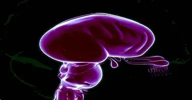
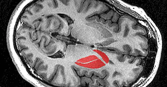
The striated body It is also known as "striated nucleus" and "neoestriate" . It is a set of structures located at the subcortical level that in turn is part of the basal ganglia, involved in the regulation of intentional and automatic movements, as well as procedural learning, reinforcement and planning.
The basal ganglia are located in the prosencephalon (or anterior encephalon), below the lateral ventricles. They are formed by the caudate nucleus, the putamen, the nucleus accumbens, the olfactory tubercle, the pale globe, the black substance and part of the subthalamus.
Technically, the term "striatum" encompasses most of the basal ganglia, with the exception of the substantia nigra and the subthalamic nucleus, since in the past these structures were conceived as a functionally related whole; however, thanks to recent research we have more information about the differences between these areas.
Today we call the whole "striated" composed of the caudate nucleus, the putamen and the nucleus accumbens , which connects the two previous structures. For its part, the concept "striated body" is used above all to designate the combination of the fluted and the pale globe.
- Maybe you're interested: "Núcleo accumbens: anatomía y funciones"
Structure and connections
The fluted body is formed by two main sections: the dorsal and ventral striatum . The first includes the putamen, the pale globe and the caudate and lenticular nuclei, while the ventral striatum is formed by the nucleus accumbens and the olfactory bulb.
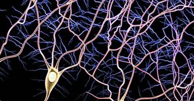
Most of the neurons that make up the striatum are medium-sized spiny neurons, which owe their name to the shape of their dendrites. We can also find Deiter neurons, which have long dendrites with few branches, and interneurons, especially cholinergic and catecholaminergic.
The caudate and the putamen, which together form the neo-striped, they receive afferents from the cerebral cortex , constituting the most important route through which information reaches the basal ganglia.
On the other hand, the eferences of the basal ganglia start above all from the pale globe, which, as we have said, forms part of the striated body according to the classical definition, but not of the striated one as such. Gabaergic emanations are sent from the pale globe (and therefore inhibitory) indirectly to the premotor cortex, responsible for voluntary movement.
Fluted functions
Together, the basal ganglia carry out very varied functions, mainly related to motor skills. These nuclei contribute to the correct functioning of the following processes:
- Motor learning
- Processing of procedural memory.
- Start of voluntary movements.
- Regulation of voluntary movements : direction, intensity, amplitude ...
- Execution of automatic movements.
- Start of eye movements.
- Regulation of working memory (or operational).
- Focus of attention .
- Regulation of motivated behavior (depending on dopamine).
- Selection of actions based on the expected reward.
The striatum is related to most of these functions, as it is the most important part of the basal ganglia. Specifically, the ventral striatum average in learning and motivated behavior through the secretion of dopamine, while the dorsal section is involved in the control of movement and in executive functions.
Related disorders
Most disorders and diseases related to the striatum affect movements, both volunteers and automatic . Parkinson's disease and Huntington's disease are two basic examples of basal ganglia dysfunction.
However, certain psychological alterations seem to be influenced by the functioning of this structure, mainly in relation to its role in the cerebral reward system.
1. Parkinson's disease
Parkinson's disease causes lesions in the brain, mainly in the basal ganglia. The death of dopaminergic neurons in the substantia nigra it interferes with the release of dopamine in the striatum, causing motor symptoms such as slowness, rigidity, tremors and postural instability. Depressive symptoms are also produced.
2. Huntington's disease
During its initial phase, Huntington's disease mainly affects the striatum; This explains why the early symptoms are related to motor control, emotions and executive functions. In this case the basal ganglia are unable to inhibit unnecessary movements , so hyperkinesia occurs.
3. Bipolar disorder
The research suggests that in some cases of bipolar disorder there are alterations in the genes that regulate the function of the striatum. Evidence in this regard has been found for both type I and type II bipolar disorder.
- Related article: "Bipolar disorder: 10 features and curiosities you did not know"
4. Obsessive-compulsive disorder and depression
Obsessive-compulsive disorder and depression, which they have a similar biological basis , have been associated with dysfunctions in the striatum. This would explain the decrease in mood that occurs in both disorders; The difficulty to inhibit movements is also relevant in OCD.
- You may be interested: "Are there several types of depression?"
5. Addictions
Dopamine is a neurotransmitter involved in the brain's reward system; the pleasant sensations that we feel when dopamine is released in the basal ganglia explain our motivation to return to seek experiences that we know are pleasurable. This explains addictions from a physiological point of view .