The 11 parts of the eye and their functions
The vision stands out among human sensory systems due to its high complexity. The structure of the eye, the main organ of sight, is a good example of this, to the point that it has come to be used as a supposedly irrefutable argument by those who defend that life was created and designed by a god.
The analysis of the parts of the eye It can be greatly extended since the organs of vision are composed of many structures. In this article we will focus on the main ones and the general description of the transduction process that makes the light energy come to be perceived as images.
- Maybe you're interested: "Nociceptors (pain receptors): definition and types"
What is the eye?
The eyes are the basis of the visual system. These organs transform light energy into electrical impulses that, when transmitted to the visual cortex of the occipital lobe, allow three-dimensional perception of shape, movement, color and depth.
The eyeballs have a spherical shape and an approximate diameter of 2.5 cm. They are divided into two sections: the anterior chamber and the posterior chamber, filled respectively with aqueous and vitreous humor, liquids that regulate intraocular pressure. The anterior chamber is smaller and lies between the cornea and the iris, while the posterior chamber is made up of the rest of the parts of the eye.
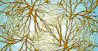
Unlike what happens with other sensory organs, the eye it is partially derived from the central nervous system . In particular, the retina, which receives the light information, develops from the diencephalon, the embryonic structure that also gives rise to the cerebral hemispheres, the thalamus and the hypothalamus.
In the retina we find Two types of photoreceptors, canes and cones . While the cones allow daytime vision and the perception of color and detail, the canes are adapted for night vision and produce low resolution images in black and white.
Parts of the eye and its functions
The eyes work in a similar way to the cameras. The lens is adjusted according to the distance of the stimulus, serving as a kind of lens that allows the refraction of light; The pupil is the diaphragm through which the image enters the eye and projects into the retina, from where it will be sent to the brain through the optic nerve.
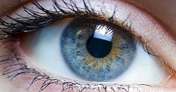
1. Cornea
The cornea is the anterior part of the eye and is in contact with the outside. It is a transparent structure that covers the iris and lens and allows light refraction . Tears and aqueous humor allow the correct functioning of the cornea, since they perform functions equivalent to those of the blood.
2. Iris
This structure separates the anterior and posterior chambers of the eye. The dilator muscle of the iris increases the size of the pupil (mydriasis) and the sphincter muscle reduces it (miosis). The tissue of the iris is pigmented because of the presence of melanin ; this gives rise to the color of the eye, by which we can easily identify this structure.
3. Pupil
There is a circular hole in the center of the iris that allows regulate the amount of light that enters the eye when changing size as a result of mydriasis and myosis; This opening is the pupil, the dark part that is located in the center of the iris.
4. Crystal
The lens is the "lens" that sits behind the iris and allows visual focus. Accommodation is the process by which the curvature and thickness of the lens are modified to Focus objects according to their distance . When the rays of light pass through the lens, the image is formed in the retina.
5. Watery humor
The aqueous humor is found in the anterior chamber of the eyeball, between the cornea and the lens. Nourish these two structures and allows eye pressure to remain constant . This liquid is composed of water, glucose, vitamin C, proteins and lactic acid.
6. Sclera
The sclera covers the eyeball, giving it its characteristic white color and protecting the internal structures. The anterior part of the sclera is attached to the cornea, while the posterior part has an opening that allows the connection between the optic nerve and the retina.
7. Conjuntiva
This membrane coats the sclera. Contributes to the lubrication and disinfection of the eyeball since it produces tears and mucus, although the lacrimal glands are more relevant in this regard.
8. Choroids
We call "choroidal" to the layer of blood vessels and connective tissue that separates the retina and the sclera.The choroid provides the retina with the nutrients and oxygen it needs to function properly, in addition to maintaining a constant temperature in the eye.
9. Vitreous humor
The posterior chamber of the eye, which is situated between the lens and the retina, is full of vitreous humor, a gelatinous liquid of greater density than that of aqueous humor from the previous camera. It is the largest part of the eyeball and has the functions of stiffening, cushioning impacts, maintaining intraocular pressure and fixing the retina.
10. Retina
The retina is the true receiving organ of the visual system since in this structure the rods and cones, the photoreceptor cells are located. This membrane covers the back of the eye and has a function similar to that of a screen: the lens projects the images perceived in the retina, from where it will be transmitted to the brain through the optic nerve.
Specifically, light rays are received by the area of the retina known as fovea , that being very rich in cones has a great visual acuity and therefore is the main one in charge of the detail vision.
11. Optic nerve
The optic nerve is the second of the twelve cranial nerves. It is a set of fibers that transmit the light impulses from the retina to the cerebral optic chiasm . From this point, visual information is sent to other areas of the brain in the form of electrical signals.
- Related article: "Cranial pairs: the 12 nerves that leave the brain"