The 5 main technologies for the study of the brain
The human brain is a mystery, but it is, too, one of the mysteries that have generated most interest throughout history .
After all, millennia ago it is known that it is there where thoughts, feelings, subjective sensations and self-awareness arise. In addition, this set of organs is so complex that until recently who wanted to study it could only do so passively and indirectly, ie, examine the brains of people already deceased and try to relate the symptoms expressed by this person with the anatomy of their nervous organs.
With what technologies is the brain and nervous system studied?
This had clear drawbacks: you could not contrast this type of information with what was observed in the behavior of the person in real time (which meant among other things that you could not get useful data for the treatment of patients), nor could one directly study brain activity, only present in living people. The latter is very relevant, taking into account that the brain is being formed in part by the activity that is in it: the characteristics of the nervous functioning dynamics of each one modify the anatomy of the brain .
Fortunately. today there are technologies that allow to study not only the anatomy of the brain of living and conscious people , but also its operation and activity in real time. These new techniques are enceplography (EGG), computerized axial tomography (CAT), positron emission tomography (or PET), angiogram and functional magnetic resonance imaging (fRMI). Next we will see the characteristics of each one of these systems.
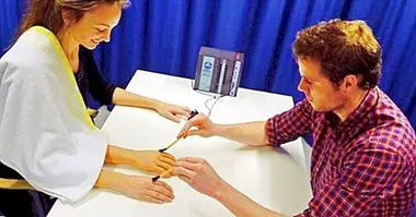
1. Electroencephalography, or EEG
This has been one of the first methods developed to "read" the activity of the brain, that is, the electric firing patterns that run through it. The technique is relatively simple, and consists of leaving fixed electrodes on the scalp of the person so that they capture the electrical impulses that capture just below to send this information to a machine. The machine collects this data and expresses it in the form of lines and peaks of activity by means of a graphic plotter, in the same way as seismographs that measure the intensity of earthquakes work. This activity record is called encephalogram .
The EEG is very simple and versatile, so it can be used to measure the activity of a few neurons or larger areas of the cerebral cortex. It is widely used to study cases of epilepsy, as well as the brain waves of sleep, but as it is not very precise it does not allow us to know exactly in which part of the brain these activation patterns are initiated. In addition, knowing how to interpret encephalographies is complicated and you need a good training and training to do so.
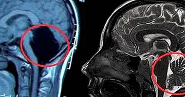
2. Computerized axial tomography, or CAT scan
The computed axial tomography (CAT) , unlike encephalography, it gives us an image of the brain and its anatomy seen from various angles, but not of its activity. That's why it basically serves to study the forms and proportions of different parts of the brain at any given time.
3. Positron emission tomography, or PET
This type of tomography it does serve to study brain activity in specific areas of the brain, albeit indirectly. To apply this technique, a slightly radioactive substance is injected into the person's blood, which will leave a trace of radiation where it passes. Then, some sensors will detect in real time, which areas of the brain are those that monopolize a greater radiation, which may indicate that these areas are absorbing more blood because, precisely, they are being more active.
From this information a screen is recreated the image of a brain with the most activated areas indicated .
4. Angiogram
The angiogram It looks a bit like PET, although in this case a kind of ink is injected into the blood. In addition, the ink is not accumulated for a while in the most activated areas of the brain, contrary to what happens with radiation, and it keeps circulating through the blood vessels until it disappears, so it does not allow to obtain an image of the brain. brain activity and yes of its structure and anatomy.
It is specially used to detect areas of the brain that are diseased .
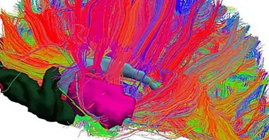
5. Magnetic resonance imaging (MRI and fMRI)
Both the magnetic resonance imaging as its "extended" version, functional magnetic resonance imaging or fMRI, are two of the most popular brain study techniques in research related to psychology and neurosciences.
Its operation is based on the use of radio waves in a magnetic field in which the head of the person in question is introduced .
The limitations of these techniques
The use of these technologies is not free of inconveniences . The most obvious is its cost: the machines that are required for its use are very expensive, and to that we must add the opportunity cost of having a space reserved for a clinic and having at least one highly qualified person to lead the process.
In addition, the information related to the parts of the brain that are activated do not always provide much information, since each brain is unique. That makes the fact that a part of the cerebral cortex "lights up" does not have to mean that the part responsible for X function has been activated.