The 7 types of neurological tests
The nervous system in a set of organs and structures, formed by nervous tissue, which are responsible for collecting and processing signals to then control and organize the other organs, and thus get a correct interaction of the person with their environment.
The science responsible for studying all this complex structure is neurology. Which tries to evaluate, diagnose and treat all kinds of disorders of the nervous system. For the work of evaluation and diagnosis a series of neurological tests have been developed that allow medical personnel to observe the operation of said system.
- Related article: "The 15 most common neurological disorders"
What are neurological tests?
Neurological tests or examinations are performed in order to examine whether the patient's nervous system is functioning properly. These tests can be more or less exhaustive depending on what the doctor tries to assess, in addition to the age or the state in which the patient is.
The importance of these tests lies in their usefulness in detecting possible alterations early , and thus eliminate or reduce, as far as possible, possible complications that may appear in the long term.
The first tests that the clinician performs are the physical tests, in which by means of the use of hammers, tuning forks, flashlights, etc. the nervous system is put to the test.
The aspects that are evaluated during this type of neurological examination are:
- Mental state (consciousness)
- Reflexes
- Motor abilities
- Sensory capacities
- Balance
- Operation of the nerves
- Coordination
However, in the case that there is suspicion of a possible alteration in any of these aspects, the medical professional has at his disposal a large number of specific and very revealing clinical tests at the time of diagnosing any type of neurological problem.
Types of neurological tests
There are more than a dozen tests to evaluate the state of the nervous system, any of them will be more or less useful depending on what the clinician wants to look for.
Here some of them are explained.
1. Brain angiography
Cerebral angiography, also known as arteriography, is a procedure to locate possible vascular singularities in the brain . These irregularities range from possible brain aneurysms, obstructions of the blood vessels or stroke, to brain inflammations or malformations in the veins of the brain.
To detect any of these abnormalities, the doctor injects a radiopaque substance into one of the cerebral arteries, thus making visible any vascular problems in the brain on the radiographs.
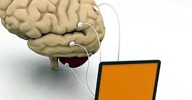
2. Electroencephalogram (EEG)
If what the doctor needs is to monitor brain activity, the electroencephalogram can be his reference test. During this test a series of electrodes are placed on the patient's head, these small electrodes transport the electrical activity of the brain to an apparatus that reads said activity and converts it into a trace of the electrical record.
Likewise, the patient may be subjected to various tests in which he is presented with a series of stimuli such as lights, noise or even medication . In this way the EEG can detect changes in brain wave patterns.
If the medical professional sees it necessary to further narrow the search or make it more exhaustive, it may be possible to place these electrodes directly in the patient's brain through a surgical incision in the patient's skull.
The electroencephalogram is very interesting when diagnosing diseases or alterations such as
- Brain tumors
- Psychiatric disorders
- Metabolic disorders
- Injuries
- Brain or spinal inflammation
- Seizure disorders
3. Lumbar puncture
Lumbar punctures are performed with the objective of obtaining samples of cerebrospinal fluid . This fluid is analyzed to check for bleeding or brain hemorrhages, as well as to measure intracranial pressure. The aim is to diagnose a possible infection of the brain or marrow, such as those that occur in some neurological diseases such as multiple sclerosis or meningitis.
Commonly, the procedure to follow in this test begins by laying the patient on one side, asking him to place the knees next to his chest. The doctor then places the position between the vertebrae in the middle of which the puncture will be performed. After administering a local anesthetic, the doctor inserts a special needle and extracts a small sample of fluid.
4. Computerized tomography (CT)
This test is part of the so-called brain ultrasound , among which are also magnetic resonance and positron emission tomography. The advantage of all of them is that they are painless and non-invasive processes.
Thanks to computerized tomography, quick and clear images of organs, brain, tissues and bones are obtained.
Neurological CT can help to make differential diagnoses in neurological disorders with several similar properties. In addition, it is especially effective in detecting, among others:
- Epilepsy
- Encephalitis
- Clots or intracranial bleeds
- Brain damage due to injury
- Brain tumors and cysts
The test lasts around 20 minutes, during which the patient must remain rested inside the CT chamber. For this test the person must remain very still while X-rays scan their body from different angles.
The final result is several transverse images of the internal structure, in this case the internal structure of the brain. Occasionally, contrast fluid can be introduced into the bloodstream to facilitate the differentiation of different brain tissues.
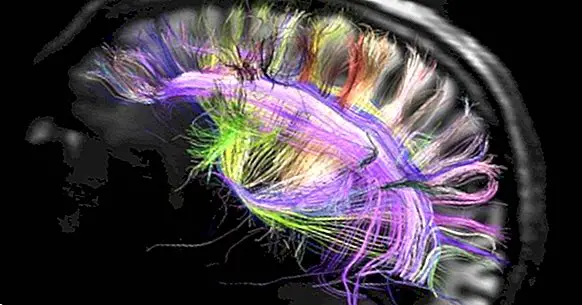
5. Magnetic resonance (MR)
To obtain images obtained by magnetic resonance, radio waves are used that are generated in an apparatus and a large magnetic field that reveal the details of organs, tissues, nerves and bones.
As in CT, the patient must remain reclining and immobile and to which is inserted inside a hollow conduit surrounded by a large magnet.
During the test, a large magnetic field is created around the patient and through a series of reactions a resonance signal is produced from various angles of the patient's body. A specialized computer treats this resonance by converting it into a three-dimensional image or a two-dimensional transverse image.
Also, there is also functional magnetic resonance, in which images of the blood flow of different areas of the brain are obtained thanks to the magnetic properties of the blood.
6. Positron emission tomography (PET)
In positron emission tomography the clinician can get images, in two or three dimensions, of brain activity . This image is achieved through the measurement of radioactive isotopes injected into the patient's bloodstream.
These radioactive isotopes attached to chemicals that run to the brain are tracked while the brain performs different tasks. Meanwhile, gamma-ray sensors scan the patient and a computer processes all the information by displaying it on a screen. Different compounds can be injected to examine more than one brain function at a time.
PETs are especially useful when it comes to:
- Detect infected tumors and tissues
- Determine brain changes after the consumption of substances or injuries
- Evaluate patients with memory disorders
- Seizure disorders evaluation
- Measure cellular metabolism
- Show blood flow
7. Evoked potentials
In the evoked potential test, possible sensory nervous problems can be evaluated , as well as corroborate certain neurological conditions such as brain tumors, marrow lesions or multiple sclerosis.
These potentials or evoked responses calibrate the electrical signals that visual, auditory or tactile stimuli send to the brain.
Through the use of electrode needles, nerve damage is evaluated. A pair of these electrodes measures the electrophysiological response of the stimuli in the patient's scalp, and the other pair is placed in the area of the body that is to be examined. Next, the clinician notes the time it takes for the impulse generated to reach the brain.
Other tests frequently used for the evaluation and diagnosis of neuronal disorders are:
- Biopsy
- Single photon emission tomography
- Doppler ultrasound
- Myelography
- Electromyography