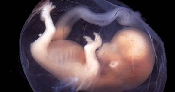
The development of the nervous system during pregnancy
The development of the nervous system begins at the beginning of pregnancy . Initially neurons are undifferentiated cells of any other, but the interaction of various factors causes them to evolve and form an elaborate tissue of synaptic connections that will allow the coordination of the functions of the organism.
Let's see what this process consists of and what are the main phases of the formation of the system in the prenatal stage of the life of a human being.
- Related article: "The 3 phases of intrauterine or prenatal development: from the zygote to the fetus"
The formation of the nervous system
Fecundation consists of the penetration of a sperm into the ovum after reaching the fallopian tubes. Although initially the two gametes form a single cell (the zygote) , during the first days of pregnancy it is divided successively, giving rise to a set of cells that is called a morula.
When the zygote is implanted in the uterus the division of its cells begins to give rise to the embryo and the placenta; during this period we refer to the embryo as "blastula". This moment supposes the beginning of the cellular differentiation.
In the initial weeks of pregnancy, the embryo is formed by three layers of cells, which are respectively called endoderm, mesoderm and ectoderm. Throughout the intrauterine development the body will be formed from these cellular assemblies.
The endoderm layer progressively becomes the respiratory and digestive tracts, while the mesoderm gives rise to the bones, muscles, circulatory system and notochord, from which the spinal column develops. The nervous system and the skin arise from the ectoderm , the outermost layer of the three.
- Related article: "How to take care during the first month of pregnancy: 9 tips"
The development of the neural tube
During the first weeks the ectoderm evolves to become the flat oval plate. This plate has a groove, the neural groove, which will give rise to the neural tube when joining the segments of the plate.
The peripheral nervous system appears from the neural crests, portions of the oval plaque that separate from it when the neural tube closes. The neural tube will later become the medullary canal and in the cerebral ventricles; from its walls the central nervous system will emerge.
Towards the end of the first month of gestation the anterior part of the neural plate is divided into three sections that shortly after will form the encephalon: the forebrain will become the cerebral cortex, the thalamus, the hypothalamus and the basal ganglia, the mesencephalon in the brain stem and the rhombencephalon in the cerebellum, the pons and the medulla.
Proliferation, migration and neuronal differentiation
On the inner side of the wall of the neural tube, the ventricular zone is located, where cell proliferation occurs. This phenomenon, which will continue until birth, consists of the production of large amounts of nerve cells (neurogenesis) through successive mitoses or cell divisions.
At this point the neural cells are still undifferentiated. Although many will remain in the neural tube for the moment and will be transformed into neurons later, others will become glial cells and move to other regions.
Neuronal migration consists in the movement of neuroblasts , primal neural cells very similar to "stem cells", from the ventricular zone of the neural tube to their respective destinations in other parts of the brain. Radial glia allows migration since future neurons move through their extensions.
Upon reaching their final position, the neuroblasts begin to transform into different types of neurons depending on the genetic information they contain, the area in which they are located and the neurons around them (which is known as "induction") ; this process is cell differentiation.
Synaptogenesis, apoptosis and reorganization
The dendrites and axons of the neurons have extensions, the growth cones, which adhere to surfaces in order to favor the growth of the neuron. In this process neurotrophic factors intervene , chemical substances that when released by neurons attract or repel axons.
When the axons reach their destination they begin to branch out, connecting with other nearby cells; thus synaptogenesis or synapse formation begins, which will develop definitively after birth, thanks to the influences of learning.
During the initial neuronal proliferation and synaptogenesis an excessive number of neurons and synapses are formed, which nevertheless allows all the basic connections to take place. Once these processes have been completed apoptosis or programmed neuronal death occurs , which causes between 20 and 80% to degrade to death.
Apoptosis mainly affects the most "weak" neurons, that is, those that have not synaphed with other cells or that have not been attracted by neurotrophic factors. This keeps only the most efficient and solid connections.
After the neuronal death the synapses are reorganized: some of the connections that were established are canceled and new ones appear until a complex and highly interconnected neural network is established that will continue to evolve and perfecting itself during growth.
- Related article: "Sinaptogénesis: how are connections created between neurons?"
Myelination and nerve conduction
In the fourth month of gestation the glial cells begin to form myelin sheaths around the axons. This substance increases the speed of transmission of nerve impulses, in addition to protecting the axons.
Myelination begins in the peripheral nervous system . Subsequently it occurs in the upper part of the spinal cord, from where it spreads to the lower and upper sections of the future body.
Nerves related to motor skills are myelinated before those associated with sensation; this is why babies are born with basic reflexes. The process of myelination will intensify during the first months after birth and continue later, at least until puberty.