Types of major cells of the human body
The human body is made up of 37 trillion cells , which are the unity of life.
It is not surprising that we find a great diversification between them in order to carry out different functions, allowing us to complement and cover the vital needs of an organism, such as the maintenance of body structure, nutrition and breathing. It is estimated that There are about 200 types of cells that we can distinguish in the organism, some more studied than others.
Throughout this article we will talk about the main categories that group types of cells according to their characteristics.
Why do these microscopic bodies matter?
Although our mental processes seem to arise from some hidden corner of our head in which the connection between the soul and the body is established, as the philosopher Descartes believed, the truth is that they are basically explained by the relationship between the human organism and the human body. the environment in which he lives. That is why knowing the types of cells we are composed of helps us understand how we are and in what way we experience things.
As you can imagine, we will not talk about each of them, but we will do some general brushstrokes about some of them to better understand our body.

Classifying cell classes
Before starting, it would be ideal to group cell types to better organize the topic. There are several criteria to distinguish different types of cells .
For the case that touches us (cells of the human being) we can classify them depending on the group of cells to which they belong, that is, in what type of tissue they can be found.
The human body is made up of four different types of tissue, thanks to which we are able to keep the different environments relatively isolated from each other that our body needs to function properly . These tissue categories are the following:
- Epithelial tissue : configures the superficial layers of the organism. In turn, it can be divided between coating and glandular.
- Conjunctive tissue : acts as a connection between tissues and shapes the structure of the body. Bone, cartilage and blood are the most specialized tissues of the conjunctiva.
- Muscle tissue As its name suggests, it is made up of the group of cells that make up the muscles.
- Nervous tissue : formed by all the elements that form the nervous system.
1. Epithelial tissue cells
In this group we find the cells that are part of the most superficial layers of the organism. It is subdivided into two types that we will see below with their fundamental characteristics.
1.1. Coating fabric
They are the proper layers that cover the organism.
Cells of the epidermis or keratinous : cells that make up the skin. They are placed in a compact form and are kept tightly joined together, so as not to allow the entry of external agents. They are rich in keratin fiber, which kills them as they ascend to the most superficial part of the skin, so that when they reach the outside they are hard, dry and strongly compacted.
- Pigmented cells : this type of cells is the one that gives color to the skin thanks to the production of melanin, which serves as a protector against solar radiation. The problems in these cells can cause many problems in the skin and in the vision, for example, as it happens in certain types of albinism.
- Merkel cells : these cells are responsible for providing us the sense of touch. They are interconnected with the nervous system to transmit this information in the direction of the brain.
- Pneumocytes : located in the pulmonary alveoli, have the function of bridging the air collected in the lungs with blood, to exchange oxygen (O2) for carbon dioxide (CO2). In this way, they are at the beginning of the sequence of functions responsible for bringing oxygen to all parts of the body.
- Papillary cells : cells that are found in the tongue. They are what allow us to have a sense of taste, thanks to the ability to receive chemical substances and transform this information into nerve signals, which constitute the taste.
- Enterocytes : cells of the smooth intestine, which are responsible for absorbing the digested nutrients and transmitting them to the blood to be transported. Its function is, therefore, to make the wall function permeable to certain nutrients and impassable for other substances.
- Endothelial cells : they are those that configure and structure the blood capillaries, allowing the correct circulation of the blood.Failures in these cells can cause cell damage in very important organs, which would stop functioning properly and, in some cases, this can lead to death.
- Gametos : are the cells that participate in the fertilization and formation of the embryo. In the woman it is the ovule and in the man it is the sperm. They are the only cells that contain only half of our genetic code.
1.2. Glandular tissue
Groups of cells that share the function of generating and releasing substances.
- Sweat gland cells : types of cells that produce and expel sweat to the outside, mainly as a measure to reduce body temperature.
- Lacrimal gland cells : they are responsible for generating the tear, but they do not store it. Its main function is to lubricate the eyelid and make it slide correctly above the eyeball.
- Salivary gland cells : responsible for producing saliva, which facilitates the digestion of food and, at the same time, are a good germicidal agent.
- Hepatocytes : belonging to the liver, perform several functions, including the production of bile and the energy reserve of glycogen.
- Calciform cells : cells found in various parts of the body, such as the digestive or respiratory system, which are responsible for generating the "mucus", a substance that serves as a protective barrier.
- Palietal cells : located in the stomach, this class of cells are responsible for producing hydrochloric acid (HCl), responsible for the proper production of digestion.
2. Connective tissue cells
In this category we will find the types of cells that are part of the body's structural and connective tissue.
- Fibroblasts : they are large cells that are responsible for the maintenance of the entire body structure thanks to the production of collagen.
- Macrophages : types of cells that are found around the periphery of connective tissue, especially in areas with a high risk of invasion, such as in the entrance to the body, with the function of phagocytizing foreign bodies and presenting antigens.
- Limfocitos Commonly grouped in leukocytes or white blood cells, these cells interact with the antigens indicated by the macrophages and are responsible for generating a defense response against it. They are the ones that generate the antibodies. They are divided into type T and B.
- Monocytes : they constitute the initial form of the macrophages but, unlike these, circulate through the blood and are not seated in a specific place.
- Eosinophils : they are a class of leukocytes that generate and reserve different substances that are used to defend against a parasitic invasion by a multicellular organism.
- Basophiles : white blood cells that synthesize and store substances that favor the inflammation process, such as histamine and heparin. Responsible for the formation of edemas.
- Mast cells : class of cells that produce and reserve large quantities of substances (including histamine and heparin) that release them as a defensive response, helping the other cells of the immune system.
- Adipocytes : cells that are found throughout the body and have the ability to capture fats as energy reserves, mainly.
- Chondroblast and chondrocytes : they are in charge of forming the tissue that we know as cartilage. Chondroblasts produce chondrocytes, which have the function of producing the necessary components to form cartilage.
- Osteoblasts and Osteocytes : cells responsible for forming bones, generating the process of calcification and therefore conditioning the process of growth and maturation of people. The difference between the two is that the osteoblast is the initial phase of an osteocyte.
- Red blood cells : also known as erythrocytes, this type of cell is the main one in the blood, transporting the O2 to the cells and extracting the CO2 to the lungs. They are the ones who give the distinctive color of blood by containing the protein hemoglobin.
- Platelets or thrombocytes : small cells that are activated when a blood vessel has been damaged and it is necessary to repair it to avoid blood loss.
3. Muscle tissue cells
In this group we only find a single type of cell that structures the muscles, responsible for the mobility of the organism.
- Of muscle fibers or myocytes : the main cell that shapes the muscles. They are elongated and have the ability to contract. The muscle fibers can be differentiated between striated skeletal, which allows us voluntary control of the body; striated cardiac, not voluntary and is responsible for keeping the heart moving; and smooth, involuntary nature that controls the activity of other internal organs, such as the stomach.
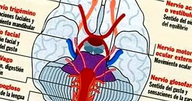
4. Cells of nervous tissue
Finally, in this category are the cells that are part of the nervous system.
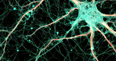
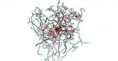
- Neurons This type of cell is the main one of the nervous system, which has the function of receiving, driving and transmitting nerve impulses.
- To further expand on the subject, you can read the article "Types of neurons: characteristics and functions".
- Neuroglia : set of cells with the function of supporting neurons, such as protection, isolation or means through which to move, mainly.
- Cones : cells found in the retina, which capture high-intensity light, providing the sense of daytime vision. They also allow us to differentiate colors.
- Canes : cells that work together with the previous ones in the retina, but capture the light of low intensity. They are responsible for night vision.