Cerebral peduncles: functions, structure and anatomy
The human brain it is such a complex organ that in order to function properly it must have the participation of a large number of parts and structures in its anatomy. Several of these parts of the brain are large and easy to distinguish with the naked eye, such as the frontal lobes, but others are very small and are located below these folds of the surface.
This is the case of the cerebral peduncles , small regions of the brain that, despite their size, are very important. Then you can learn more about these anatomical structures.
What are the cerebral peduncles?
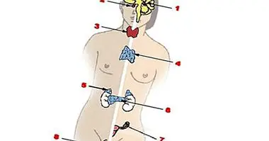
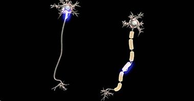
The human brain is composed, broadly speaking, by two main types of "material": gray matter and white matter. The first is the one that contains a higher concentration of neuronal soma "that is, of" bodies "of neurons, the zone in which the nucleus of these cells is located), while in the areas with white matter, other anatomical areas predominate. the neuron: the axons, narrow and elongated elements that when grouped in the form of bundles form nerve fibers.
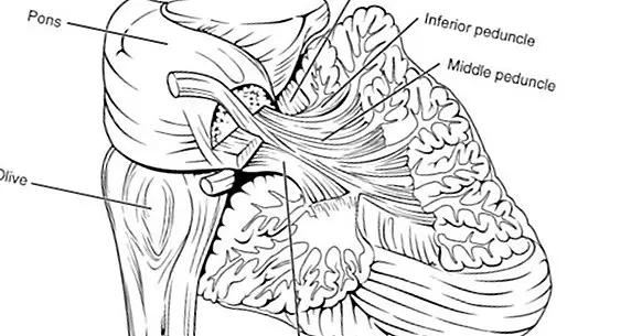
The cerebral peduncles are two small structures of cylindrical shape and white color which are composed of white matter. They are born from the brainstem, specifically above the Varolio bridge, and reach the cerebral cortex projecting vertically.
Between the peduncles of the brain is the interpeduncular fossa, a space that acts as a physical separation between these two structures as if it were a kind of tunnel. Further, the ventral and dorsal side of each peduncle are separated each other by a sheet of something that is not white matter, but something known as black substance.
They are not the same as the cerebellar peduncles
It is easy to believe that the cerebral peduncles are the same as the cerebellar peduncles, but this is not the case. These second structures have a similar function , since they also function as important nerve pathways that collect information from various regions. However, both their location and the type of information that make up these neural signal channels are different.
Functions of the cerebral peduncles
The cerebral peduncles are fundamentally "autopiestas" of nervous impulses that travel through the brain. As they are made of white matter, this information goes through them quickly, since the myelin that covers the axons (and that gives that white tone to these structures) makes these electrical signals go at a great speed.
The main function of the cerebral peduncles, then, is that of communicate two areas of the brain : the mesencephalon (located in the upper part of the brainstem) and the cerebral cortex. In fact, these nerve fiber conduits reach the division that is created between the two cerebral hemispheres.
In fact, peduncles contain several pathways. One of these is the corticospinal, which, as its name indicates, communicates the cortex with the spinal cord, and the other is the corticopontine tract , which joins the bark with the Varolio bridge.
As the brainstem is responsible for executing automatic and stereotypical actions useful or necessary for our survival, the role of the cerebral peduncles also has to do with this task.
Thus, the main functions of these bundles of nerve fibers are the regulation of motor impulses and the transmission of reflex acts, two very useful mechanisms to keep us alive, since they allow us to react quickly to significant events (especially if they are dangerous or harmful).
Specifically, peduncles have an important role in the reflex movements of the eyes , necessary for them to work well, and in the coordination of these acts with other movements of the head and neck.
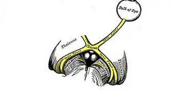
Specifically, these structures intervene in the reflex control of reflex movements. Normally this occurs when the orders of the motor cortex reach the cerebral peduncles, and from these they go to several nuclei of a structure known as the thalamus, located in the depths of the brain, in an area known as the diencephalon.
Parts of this pair of structures
The cerebral peduncles receive connections of the cerebellum, the cerebral cortex and the lower areas of the brainstem and spinal cord.
On the other hand, the cerebral peduncles are composed of several parts or sections . One of them is the tegmentum of the mesencephalon, to which the cerebral crus happens, and the other region is called pretectum. The tectum extends beyond the brainstem, reaching the cortex of the brain.
The complexity of the different components of the peduncles has to do with the fact that these are in an area of the central nervous system in which all kinds of functions of control and coordination of specialized actions are carried out and created by millions of people. years of evolution and natural selection. In little space, the brainstem presents tiny areas responsible for performing such vital actions as the regulation of the heartbeat or the control of body temperature.