Ectoderm: what it is and how it develops during pregnancy
The biggest system or organ that shapes us, both humans and animals, is the skin. Said organ fulfills the function of protective barrier of the whole organism and is composed of three main layers: the epidermis, the hypodermis and the hypodermis. The first one, the epidermis (the outermost layer of the skin), begins its development from the embryonic period, from an earlier set of tissues that is called ectoderm .
In this article we will see what is and what the ectoderm is responsible for, as well as the specific moment of the development in which it originates.
- Related article: "The 3 phases of intrauterine or prenatal development: from the zygote to the fetus"
What is ectoderm?
The ectoderm is the outer germ layer in the early embryo . It is one of the three germ layers of embryonic origin, found in both vertebrate animals and invertebrate animals. Broadly speaking, it is a group of cells that make up the great tissues of our body, and that arise from the first weeks of gestation.
The ectoderm has been studied since 1817, when Christian Pander, a PhD student at the University of Würzburg, Germany, discovered two embryonic plates in vertebrates, which later led him to discover a third which was later called ectoderm. Subsequently, in 1825, the embryologist Martin Rathke discovered the same cell layers in invertebrate animals .
Towards the 19th century it was Karl Ernst von Baer of the University of Konigsberg in Prussia, who extended these investigations and took them to different species. The first researcher is attributed the first description of the stage of the blastula, which we will see developed later.
How does it develop in pregnancy?
During embryonic development, the cells undergo a multiple process of cell division. Eventually, the cells generated by this process reach a stage called gastrulation . It is in the latter when the embryo organizes three different germ layers.
One of these layers is the ectoderm. The others are the mesoderm and the endoderm. Together, the three layers that make up the tissues of the skin, nerves, organs and muscles. They differ from each other in the depth to which they are found, as well as in their particular functions.
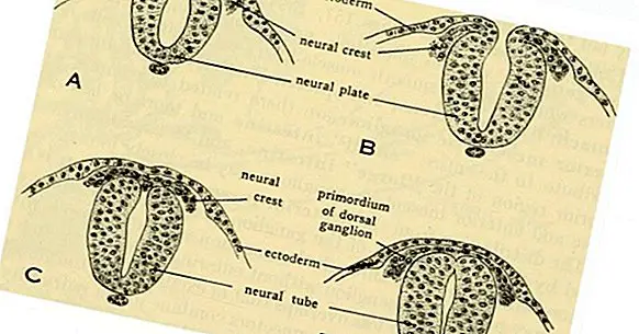
Once the gastrulation is completed, the embryo enters another stage known as neurulation, at which point the nervous system begins to develop. This stage is characterized by a thickening of the ectoderm, which allows generating "neural plates". In turn, the neural plates gradually thicken and lay the foundations of both the development of the nervous system .
In other words, the central nervous system is formed from a first neural plate composed of ectodermal cells that are found on the dorsal surface of the embryo. This generates a neural tube that will later form the ventricles and the cells necessary to consolidate the peripheral nervous system and the motor fibers that compose it. To better explain this process, the ectoderm has been divided into different parts.
- Maybe you're interested: "Endoderm: parts and development in pregnancy"
Parts of the ectoderm
During the stage of neurulation, the ectoderm is divided into two large parts : the superficial ectoderm and the neuroectoderm.
1. Surface ectoderm
The superficial ectoderm gives rise to tissues that are on the outer surface of the body , for example the epidermis, hair or nails.
2. Neuroectoderm
In neuroectoderm, it is divided into two main elements, which will later shape the nervous system. One of them is the neural tube, precursor of the central nervous system in the embryo, as well as the brain and spinal cord.
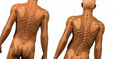
The other one is the neural crest , which gives shape to many of the bones and connective tissues of the head and face, as well as parts of the peripheral nervous system, such as some nerve ganglia, and also the adrenal glands and melanocytes (those that give rise to myelin).
In other species, the ectoderm fulfills similar functions. Specifically in fish, the neural crest shapes the spine, and in turtles it helps to form the carapace.
Its functions
As we have seen, the ectoderm is the layer from which the skin and all the sensitive structures derive . Being a layer, it consists of groups of cells that fuse with each other during the embryonic development of all animals. In vertebrate animals, the ectoderm is responsible for the development of the following tissues:
- Skin
- Nail
- Eye lens
- Epithelium , that is, the tissue that covers the organs that regulate the senses.
- Scalp and hair
- Nasal Cavity
- Paranasal sinuses
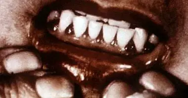
- Mouth, including tooth enamel
- Anal channel
- Nervous tissue , including endocrine cells such as the pituitary body and the chromaffin tissue
On the other hand, in invertebrate animals such as cnidarians or ctenophores (relatively simple aquatic animals of the taxonomic category "phyla"), the ectoderm covers the entire body, so in these cases the epidermis and the ectoderm is the same layer.
Bibliographic references:
- Ectoderm (2018). Encyclopedia Britannica. Retrieved August 22. Available at //www.britannica.com/science/ectoderm.
- MacCord, K. (2013). Ectoderm. The Embryo Project Encyclopedia. Retrieved August 22, 2018. Available at //embryo.asu.edu/pages/ectoderm.
- Martos, C. (2018). Ectoderm: Parts, Derivatives and Alterations. Lifeder.com Retrieved August 22, 2018. Available at //www.lifeder.com/ectodermo/.
- Poch, M.L. (2001). Neurobiology of early development. Educational Contexts, 4: 79-94.