Motor neurons: definition, types and pathologies
Our brain controls and allows our movements. Although this may seem a very reductionist description, it nevertheless remains real. Our nervous system, within which is the brain, is responsible for sending signals to all the muscles of our body so that they move.
To be more exact, these signals are sent by motor neurons or motor neurons . Thanks to which we can walk, breathe, sneeze or get our heart beat.
- Related article: "Types of neurons: characteristics and functions"
What are motor neurons?
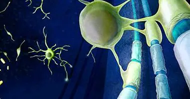
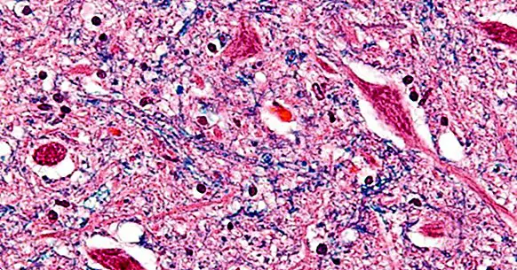
Motor neurons, also known as motoneurons, are a group of neurons in the central nervous system whose main mission is to send a series of nerve impulses to the muscles or glands. These neurons are found in the brains of all vertebrate species . In the human species, if they place especially in the spinal cord and in area 4 of Brodman.
Motor neurons are considered efferent neurons, since they are responsible for sending information from these regions to the rest of the muscles of the body; unlike afferent or sensory neurons that perform the opposite route, sending information from the muscles to the rest of the nervous system.
This transmission of nerve impulses is intended to control the skeletal muscles and smooth muscles that make up the organs and glands. That is, thanks to motor neurons we are able to perform any type of movement, just as our organs are able to function correctly.
However, in order to carry out these functions, motor neurons need the information sent by sensory or efferent neurons. Since to be able perform the muscle movements appropriate to the situation , our brain must receive information from outside. Hence the need for both types of neurons to work in consonance.
In this way, our nervous system integrates information from both types of neurons and allows us to move and react according to the demands and circumstances of our external context.
Although motor neurons have traditionally been considered passive channels of information transmission, some results obtained in recent studies point to the idea that these nerve cells have much more complex operating dynamics , being able to produce motor behaviors or patterns by themselves.
- You may be interested: "Via afferent and via efferent: types of nerve fibers"
Motoneurons and motor units
Since each neuron aims to activate a specific muscle fiber to be able to carry out a certain movement, each of these unions are called motor units. These functional units can be divided into several types:
1. Slow motor units (S or slow)
In this type of motor units, the neurons stimulate a small muscle fibers, also coined with the name red fibers, which perform very slow contraction movements.
This type of fibers tolerate tiredness and fatigue very well so they are especially apt to maintain a contraction or muscle posture without fatigue. For example, they help us to stand up without getting tired .
2. Fast fatigue motor units (FF or fast fatiguing)
In this second case, the fibers involved are the white fibers, which are responsible for innervating larger muscle groups. Compared to slow motor units, fast fatigue motor units have very short reaction times but they deplete their energies more quickly and, therefore, tire much earlier.
These motor units are extremely effective for performing movements that require rapid bursts of energy, such as jumping or running .
- Related article: "Parts of the human brain (and functions)"
3. Fast motor units resistant to fatigue
Finally, this last type of motor units are halfway between the two previous groups. Although they perform their function on medium-sized muscles, your reaction time is slower that in FF units and have the ability to tolerate fatigue for longer.
Types of motor neurons
As mentioned above, each neuron has a fundamental role in the activation of a specific fiber or tissue; so you can make a classification of different types of neurons according to the tissue on which they exert their influence.
1. Somatic motor neurons
This type of motor neurons act on the skeletal musculature, so they have a transcendental role in locomotor skills .
These skeletal muscles are formed by striated fibers, which make up most of the body mass and are distinguished from the rest by being muscles that we can move at our will.
In addition, within this group of somatic motor neurons we can find two more subgroups. The first of these subgroups serves to classify the neurons according to their position, while the second divides them according to the fibers to which they are connected.
Classification according to position
- Upper motor neuron : These neurons are located throughout the entire cerebral cortex and their nerve endings are arranged so that they form a pyramidal path connected to the spinal cord.
- Lower motor neuron In this case the neurons are arranged forming circuits, located in the anterior horn of the spinal cord, which intervene in reflex movements and involuntary movements.
Classification according to fibers
- Alpha motor neurons : they are the largest motor neurons and their main function is that of active extrafusal fibers. That is, all those fibers that make up the skeletal muscles. Thanks to them we can generate the necessary force to contract and move our muscles.
- Beta motor neurons : these neurons are connected to skeletal muscle fibers as well as fibers that are outside the muscular spindle (intrafusal) and are responsible for receiving sensory information.
- Gamma motor neurons : finally, the gamma motor neurons are only responsible for innervating the intrafusal fibers; regulating sensitivity to contraction and helping to maintain muscle tone.
2. Visceral motor neurons
The visceral motor neurons are in charge of innervating all those muscle fibers that we can not move voluntarily; that is, the smooth muscles. This musculature controls, for example, the movements of our heart, the viscera and intestines, etc.
In order to carry out its function, the visceral motor neurons also perform synapses with the neurons of the ganglia of the autonomic nervous system, sending signals to the relevant organ and innervating the visceral musculature .
3. Special visceral motor neurons
This last group of neurons has as its sole mission to activate the muscles present in the face and neck, known as the branchial musculature.
Associated pathologies
There are a number of diseases or pathologies of neurological origin that are distinguished by presenting a gradual degeneration of motor neurons, presenting a different symptomatology according to whether the affected neurons are superior or inferior .
Those diseases in which a degeneration of the upper motor neurons is experienced are characterized by a general weakening of the muscles . When the affected motor neurons are the lower ones, the person may suffer from muscular tension, stiffness and hyperactivity of the reflexes that causes involuntary muscular contractions.
Some of the diseases related to the degeneration of motor neurons are:
- Progressive bulbar paralysis.
- Pseudobulbar paralysis.
- Amyotrophic Lateral Sclerosis (THE A).
- Primary lateral sclerosis.
- Progressive muscular atrophy .
- Spinal muscular atrophy.
- Post-polio syndrome