Penfield's sensory and motor homunculi: what are they?
In the field of neurosciences they are very famous the cortical or Penfield homunculi , humanized representations of the distribution of nerves and brain structures that are related to motor and sensory functions. Different homunculi have been created for these two aspects since the cerebral topography varies between the two.
These beings look similar to people, although their members are not very proportionate; such irregularities are very useful to conceptualize the differential innervation of the parts of the body, the key aspect in the morphology of homunculi.
- Related article: "Parts of the human brain (and functions)"
What is Penfield's homunculus?
Between 1937 and 1954 the American neurosurgeon Wilder Penfield and his collaborators developed diverse representations of a striking aspect of brain topography: the presence of "maps" of nerve pathways, both sensory and motor, in the cortex.
The different functions of our organism are not represented proportionally in said map, but their size depends on the complexity of the corresponding nerves. However, the location of these brain areas does present Notable parallels with the external structure of the body .
This resulted in Penfield being inspired by the relative weight of each function in the cerebral cortex to create symbolic images of a "homunculus", a Latin term that translates as "little man" and has been used frequently throughout of history to designate artificial human beings, especially in the context of works of fiction.
Given that there are differentiated cerebral topographic representations between motor and sensory functions, we can actually find two homunculi with distinctive characteristics which is worth detailing.

What is its form?
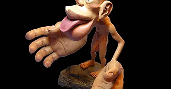
The homunculus of Penfield was described as grotesque by its own author because of the irregularity of its morphology: while hands, mouth, eyes and ears are disproportionately large in comparison with the human body, the rest of the homunculus has a weak appearance.
The comparison between the enormous hands and the arms, fragile and thin, is particularly striking. These characteristics are even more marked in the case of the motor homunculus than in the sensorial one, since the functions related to movement are less distributed than the sensory ones.
The cause of the peculiar aspect of the homunculi are the differences in the innervation of the different parts of the body : the more intense and complex the connection between one of them and the brain, the greater the size of the corresponding section in the cerebral cortex.
- Maybe you're interested: "The ghost member and the mirror box therapy"
The sensory homunculus and the somesthetic cortex
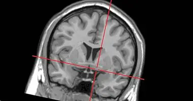
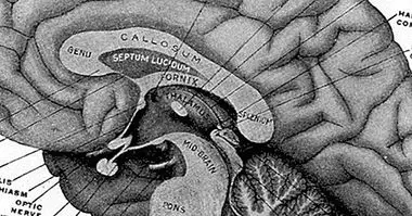
The sensory homunculus represents the primary somatic or sensory cortex , which is located in the postcentral gyrus, a cerebral gyrus located in the region of the parietal lobe attached to the frontal. In fact, Penfield was the first to describe this part of the brain, which corresponds to areas 1, 2, and 3 of the Brodmann model.
In this section of the cortex the representation of the body schema is inverted : the toes are in the upper part of the lobe, while the mouth is located in the lower part. Also the "topographic map" of each hemisphere of the body is in the opposite half of the brain. The same happens in the case of the motor homunculus.
This homunculus looks somewhat less disproportionate than the engine. However, the face and hands are very large compared to the rest of the body because these regions are endowed with many cutaneous receptors ; the density of these cells in a part of the body determines the size of their cortical representation.
The somesthetic cortex receives most of the projections of sensory information that reach the brain through the thalamus, a structure that acts as a point of connection between the cortex and other more peripheral regions.
This part of the cerebral cortex does not only deal with stimulation from the external world, but also also processes information about proprioception , that is, the sensations that the body senses about the relative position of the muscles. This sense is fundamental for movement, posture or balance, among other functions.
The motor homunculus and the primary motor cortex
The cortical representation of the motor nerves and the corresponding cutaneous receptors it is located in the primary motor cortex, in the central groove , a region of the frontal lobe that is right next to the somesthetic cortex; therefore, the two cortical homunculi are very close to each other.
The primary motor cortex is the most important area of the brain for the functioning of the motor system: it receives afferences from the thalamus and works together with the rest of regions associated with movement, such as the supplementary motor cortex, to elaborate and execute motor schemes.
The appearance of the motor homunculus is even more grotesque than that of the sensory one: its mouth, its eyes and especially its hands are enormous in comparison with the trunk, arms or legs. This is due to the greater specificity in the location of receptors and motor nerves , much less numerous than the sensory ones in a large part of the body.
Since the synaptic connections, which are the basis of the nervous system, are modified during life as a function of experience and practice, the motor homunculus changes in the same person as time passes and differs more than the sensory in the interindividual plane.