Polygon of Willis: parts and arteries that form it
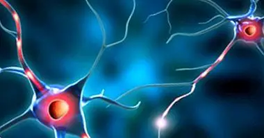
Our brain is a complex organ that governs and coordinates the totality of the systems that make up our organism. But this organ, and the nervous system in general, does not work from scratch: it needs a continuous supply of oxygen and nutrients in order to function. This contribution will reach him through the blood supply, reaching the different structures through the cerebrovascular system. Within this system we have different veins and arteries, which converge in the polygon of Willis .
- Related article: "Parts of the human brain (and functions)"
The polygon of Willis: description, location and functions
We call the polygon of Willis a structure of heptagonal form present in the brain. This structure is formed by the union of the different arteries that irrigate the brain, playing an important role in the supply of oxygen and nutrients from it. It is considered an anastomosis, or interconnection in a network of parts or elements (in this case the arteries) differentiated from each other.
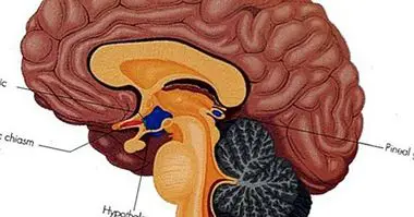
The polygon of Willis can be found in the lower part of the brain , surrounding the heptagon that forms structures such as the optic chiasm, the hypothalamus and the pituitary gland. Its structure can vary greatly from one person to another, finding that more than half of the population has a structuring of this polygon different from what is considered classic or typical.
The functions carried out by the polygon of Willis are of great importance for our survival, since through it flows the blood that irrigates a large part of the brain . In addition, we are facing the main auxiliary mechanism that allows blood to continue to reach the different regions of the brain even if there is an alteration or damage to the artery that in principle governs it. It also balances the blood supply received by both cerebral hemispheres, allowing the blood that reaches one hemisphere to communicate with the blood of others.
Arteries that converge in this polygon
As we have said, the polygon of Willis is the structure through which the different main arteries that supply the brain are interconnected. Among these arteries, the main ones and from which many others branch out are the following (although there are many other ramifications).
1. Internal carotid artery
The carotid arteries ascend through the body to the head, on both sides of the neck , to end up penetrating the skull (moment they are called internal carotids). Once inside, they will be in charge of supplying blood to the anterior part of the brain, taking care of a large part of the oxygen supply and nutrients to most of the brain (both cortex and subcortical structures), to conform with its branches the anterior part of the polygon of Willis. Later it will be divided into anterior and middle cerebral arteries, among many others.
2. Basilar artery
Another of the main arteries that supply the brain, the basilar artery, appears after the union in the brainstem of the vertebral arteries , which enter the base of the skull directly ascending around the vertebrae. This artery and its ramifications (the posterior cerebral arteries) are responsible for providing blood flow to the brainstem and the posterior regions of the brain (including the occipital lobe), forming the back of the polygon of Willis.
3. Subsequent communicating arteries
We are facing two arteries of great importance, since they allow communication between the internal carotid and the posterior cerebral artery in such a way that the main cerebral arteries on the same side of the brain are connected to each other.
4. Previous communicating artery
The anterior communicating artery is a small artery that connects the right anterior cerebral artery and the left anterior cerebral artery, acting as a bridge between both hemispheres .
5. Anterior cerebral artery
Part of the bifurcation of the internal carotid artery, this artery is part of the circle or polygon of Willis directly. Its ramifications allow to irrigate sensorimotor and orbitofrontal areas, among other areas of interest.
6. Middle cerebral artery
The larger branch of the carotid and the more vulnerable to occlusions, its blood supply tends to be directed into the brain. Your blood supply reaches the striate, the insula , and to orbital, frontal, parietal and temporal regions. It follows Silvio's fissure, which is why it is also called Silvia's or Silviana's artery.
7. Posterior cerebral artery
Artery arising from the connection between basilar artery and posterior communicating artery. Especially important for Irrigation of the lower and deeper areas of the temporal and occipital lobes , since its action allows aspects related to vision
8. Cerebellar arteries
These are the arteries that help irrigate the cerebellum, as well as other structures of the brainstem. We can find the superior cerebellum, anteroinferior and posteroinferior
9. Spinal arteries
The spinal artery is the artery that supplies blood to the spinal cord, being of great importance for the autonomic nervous system and the transmission of information from the brain to the different organs.
When injuries appear
The polygon of Willis is an area of great importance for the human being, arising in its interconnections a great amount of ramifications that can reach up to 80% of the cerebral blood flow . But sometimes you may suffer that this polygon is damaged after a trauma, that an aneurysm appears or that there is a cardiovascular accident in this region.
If some kind of obstruction appears in the polygon, it is possible that the irrigated areas run out of oxygen and die. The consequences can be multiple, from death (if for example the nuclei that regulate the vital signs are lost) to the loss of mental and physical functions, sensitivity or motor ability .
Another problem that can occur is the fact that an aneurysm appears (in fact, the polygon of Willis is one of the main places where problems of this type usually appear) and it ends up producing a spill, which can have disastrous consequences for the Affected subject. And even if the outcome is not fatal, you may lose your vision due to compression of the optic chiasm.
Bibliographic references:
- Gómez García,; .; Espejo-Saavedra, J.C .; Taravillo, B. (2012). Psychobiology CEDE Preparation Manual PIR, 12. CEDE, Madrid.
- Gray, D.J. (1985). Arterial circle of Willis. In: Treaty of Human Anatomy, Editorial Interamericana. 1st Edition: 760-3.
- Kandel, E.R .; Schwartz, J.H. & Jessell, T.M. (2001). Principles of neuroscience. Fourth edition. McGraw-Hill Interamericana. Madrid.
- Quintero-Oliveros, S.T .; Ballesteros-Acuña, L.E .; Ayala-Pimentel, J.O. and Forero-Porras, P.L. (2009). Morphological characteristics of cerebral aneurysms of the polygon of Willis: direct anatomical study. Neurosurgery, 20 (2): 110-116.