Bones of the head (skull): how many are there and what are they called?
The brain is one of the most important organs of the human body, governing the functioning of other body systems. Being protected is essential for survival.
Fortunately, we have different protection mechanisms, one of which is a strong bone covering that surrounds it. We are talking about the skull, which is composed of different bones .
- Related article: "Parts of the human brain (and functions)"
Bone protection of the brain: the skull
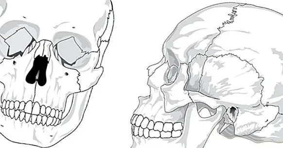
When speaking of the skull, it is usually imagined the totality of bones that are part of the head. This consideration is not entirely correct, since the skull as such is technically the bone structure that covers the brain . The rest of the bones, like those of the jaw, are part of the facial skeleton.
But nevertheless, given its use as a synonym of the set of bones of the head sometimes distinguishes between neurocranium, which would be the skull itself, which protects the brain) and viscerocranium (which would include the bone structure that gives shape to the face and which are part of the bones of the ear, the nostrils, the basin of the eyes, the nasal cavity and the set of bones that form the jaw).
In general both neurocranium and viscerocranium are solidly joined , considering that the border between one and the other marks the auditory canal and the upper part of the eye socket
The adult human skull, in its meaning as a neurocranium, is a set of eight bones welded together and bonded together throughout the development by hardened connective tissue. Its main function is the protection of the brain and allow a basic structure in which part of the facial muscles can adhere, in addition to providing a stable position to blood vessels, cranial nerves and the brain itself. Also the skull can be divided into cranial vault and base of the skull.
- Maybe you're interested: "The lobes of the brain and its different functions"
Bones that make up the skull
As we have seen, the skull or neurocranium is configured by a total of eight bones joined and welded throughout the development of the individual in what are called sutures. All of them They have different openings and holes through circulating blood vessels and nerves .
Next, the different bones that are part of the skull are exposed, as well as some of its substructures.
1. Frontal bone
This bone is placed on and protects the frontal lobe . It allows to shape the forehead and reaches to the top of the vault of the eye or supraorbital margin, being a point of union between neurocranium and viscerocranium. Its connects with the parietal bones by the coronary suture, and with the nasal bones by the frontonasal suture.
- Related article: "What is the frontal lobe and how does it work?"
2. Parietal bones
Is about the biggest bones of the skull , which form most of the upper and lateral region of this. It is connected to the frontal by the coronary suture, with the parietals by the scaly sutures and with the occipital by the lambdoid suture. Both parietals are joined together by the sagittal suture.
3. Temporary bones
Two bones each located under one of the parietal bones and joined to them by the scaly sutures. These bones, irregular, can be divided into three zones: the scaly one that is located around the scaly suture, the mastoid that refers to the part closest to the jaw in which several muscles of this and neck settle. and the stone that is located in deeper regions, forming part of the base of the skull and possessing in its interior the middle and inner ear. There is also a tympanic region , which surrounds the auditory canal.
4. Occipital bone
This bone mainly forms the base of the skull, standing in it the foramen magnum or hole in which the brain and spinal cord connect. It protects part of the occipital and temporal lobes, the cerebellum and the brainstem. It has several protuberances and ridges that connect with the vertebrae. It is connected with the parietal by the lambdoid suture and with the temporal by the occipitomastoid suture.
5. Sphenoids
This bone with the shape of a butterfly or bat It is located in an area at the height of the temple , connecting with the frontal, temporal and occipital bones. It goes from side to side of the skull, horizontally, and consists of body and major, minor, and pterygoid apophyses. In the first one you can find the Turkish chair, a structure that surrounds and protects the pituitary gland. The larger wings are part of the dorsal wall of the eye socket, while the smaller ones are part of the medial part.It holds the remaining bones of the skull together and connected.
6. Ethmoids
The bone known as ethmoids it is situated between the sphenoid and the nasal bone , participating in the formation of the ocular orbits and the nostrils, acting as the roof of the second (specifically, the part known as the lamina cribosa) and the floor of the former, as well as the separation between the two (the lateral masses are responsible for this) of the ethmoids).
This bone connects with the meninges through the crista galli. It has numerous cavities called esmoidal cells .
Bones of the viscerocranium
Although the bones of the skull are properly the previous ones, it must be taken into account that there are other bones in the structure of the head beyond them , those corresponding to the viscerocranium. In this case we can find a total of 14 bones, which together with the 8 above make up the 22 that on average has the head of an adult human (to which it is possible to add those of the ear).
Then you can see them listed, each person having two of each of the following except the vomer and the jaw (the latter being the only movable bone structure).
- Jaw
- Maxillary bones
- Nasal bones
- Lachrymal bones
- Vomer
- Cornetes
- Palatine bones
- Zygomatic bones (cheekbones)
In addition to these, inside the viscerocranium we can also find the internal ossicles of the ear that allow the reverberation of the sound up to the cluck: hammer, anvil and stirrup .
Bibliographic references:
- Rouviere, H. and Delmas, A. (2005). Human anatomy: descriptive, topographic and functional; 11th ed .; Masson