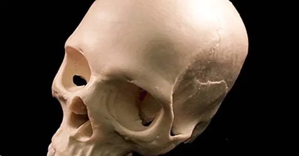
What is the human skull like and how does it develop?
Our brain is a fundamental organ for survival, given that it is the body in charge of managing and directing the functioning of other body systems, which allow us, among other things, to breathe, eat, drink, perceive the environment and interact with it.
However, its structure is relatively fragile, which requires some type of element that prevents it from being destroyed or injured by movement or falls and shocks, or that is attacked by pathogens and bacteria.
In this sense, our brain has diverse protection systems, being the most outstanding of all the bony covering that surrounds it: the human skull . And it is on this part of the body that we are going to talk about throughout this article.
- Related article: "Parts of the human brain (and functions)"
What is the human skull?
We understand by skull to the structure in the form of bone covering that surrounds and covers our brain, forming only part of what we come to consider our skull.
Its main function is to protect the whole structure of the brain, by way of barrier that prevents blows, injuries and harmful pathogens can directly attack the brain . It also allows this to maintain a structure and that there may be some buoyancy of this that prevents any blow causes it to collide with its walls, acting as a container.
While technically the skull is only the part of the skeleton that surrounds the brain (which would leave out other facial bones such as the jaw) traditionally speaking of this structure has been included along with the other bones of the facial area. In order to integrate both positions, a subdivision has been generated: the facial bones that are not part of the technical definition of the skull receive as a whole the name of viscerocranium , while the skull itself (the part that covers the brain) is called neurocranium.
Its main parts
The skull is a structure that does not appear uniformly, but is actually the union of various bones by means of cranial sutures that as we grow up end up ossifying. Between viscerocranium and neurocranium, adults have a total of 22 bones.
Among them, eight correspond and configure the neurocranium: frontal, two parietal, two temporal, sphenoid, ethmoid and occipital. All of them protect the corresponding cerebral lobes with the exception of ethmoids and sphenoids : the first of which is the structure from which the eye bones and nasal passages leave, while the second acts as a bone that binds a large part of the bones of the region and protects areas such as the pituitary gland.
The rest of the bones of the head are part of the viscerocranium, something that includes from the nostrils and lacrimal to the jaw and cheekbones.
In addition to the aforementioned bones, so-called cranial sutures are also very important in the skull. These are a type of cartilaginous and elastic tissue that join the different bones of the skull and that allow the growth and expansion of this as we develop, until finally end up becoming bone in adulthood. In this sense there are a total of thirty-seven, among which is for example the lambdoidea, the sagittal, the scaly, the spheno-ethmoidal or the coronal. Also relevant are synarthrosis or cerebral cartilage.
- Maybe you're interested: "The lobes of the brain and its different functions"
Sexual dimorphism
The skull is, as we have said, fundamental for our brain and organism, since it provides protection to our internal organs and contributes to give structure to facial physiognomy .
But not all skulls are the same. And we are not only talking about possible injuries or malformations, but there are interindividual differences and it is even possible to find differences derived from sexual dimorphism. In fact, it is possible to recognize if a skull is of a man or a woman depending on the differences between both sexes in regard to its shape and the particularities of its structure.
In general, the male skull is more robust and angled , while the female tends to be more delicate and rounded. The male skull tends to have a cranial capacity or size between 150 and 200 cc higher (although this does not imply neither greater nor lesser intellectual capacity, since this will depend on how the brain is configured, the genetic inheritance and the experiences that the subject is having in his life).
The male has a short and slightly sloping front plate, while in the female the front of the skull is smoother, bulging and high. Likewise, the temporal crest is usually very visible in the male case.
A very easy to see element are the supraorbital arcades , which are usually practically non-existent in women, whereas in men they are usually marked. The orbits are usually quadrangular and low in the man while the woman has rounded and higher.
The jaw and the teeth are very marked in the man, something less usual in the case of the woman. The chin of the woman usually is oval and little marked, whereas that of the man is very marked and is usually square. It is also observed that the occipital protuberance protrudes and is highly developed in men, something that does not happen to the same extent in women.
Training and cranial development
Like the rest of our organs, our skull is signed and developed throughout our pregnancy, although this development does not end until many years after birth.
Initially the skull it develops from the mesenchyme , one of the germinal layers that appear during embryogenesis and that arises in the fetal period (from three months of age) from the neural crest. The mesenchyme, which is a type of connective tissue, will be differentiated into different components, among which the bones will be developed (organs arise from other structures called endoderm and ectoderm).
According to our organism, the tissues are ossified. Before being born the bones of our skull are not fully formed and fixed , something that is evolutionarily beneficial because the head will be able to partially deform to pass through the birth canal.
When we are born we have a total of six cranial bones, instead of the eight that we will have as adults. These bones are separated by spaces of membranous tissue called fontanels, which will eventually form sutures that throughout the development will end up configuring the adult skull.
It will be after being born when little by little these fontanelles will be closing, beginning to take shape right after the birth (in which they return to their original position) to grow until reaching the final cranial capacity around the age of six, although the cranium will continue its growth until adulthood .
It can be said that this growth and development of the skull is often linked and produced in relation to the encephalon itself. It is mainly the cartilage and the soft tissue matrix from the bone that generate growth when expanding to try to counteract the pressure exerted by brain development, which is determined by genetic factors (although it can also be partially influenced by factors environmental).
Bone diseases and malformations
We have seen throughout the article that is the skull and how it is usually formed in most people. However, there are different diseases and situations that can cause this part of our skeleton to develop abnormally , does not close or even closes too soon (something that prevents the correct growth of the brain).
This is what happens with diseases such as Crouzon or craniosynostosis, in which, due to mutations and genetic diseases, the sutures that unite the bones close too soon.
However it is not necessary that there is a congenital problem for the skull to be deformed: in Paget's disease (the second most common bone disease after osteoporosis) occurs an inflammation of the bone tissue that can lead to deformations and fractures in the bones.
Although it is not a disease specifically of the skull (it can appear in any bone) one of the possible locations in which it can occur and where it is more frequent is precisely in it. And this may imply the appearance of complications and neurological injuries.
Other conditions such as hydrocephalus, macrocephaly, spina bifida or some encephalitis or meningitis (especially if they occur in childhood) can also affect the correct development of the human skull.
Finally, it is also worth mentioning the possibility of this happening Having suffered some traumatic brain injury , as for example in a traffic accident or an assault.
An alteration at the level of the skull can have multiple effects, since it can affect the development and functioning of the brain: it can compress and hinder the growth of the entire brain or of specific parts of the brain, it can alter the level of intracranial pressure, it can generate lesions in neural tissue or can even facilitate the arrival of infections by bacteria and viruses.
It is even possible that, even without the need for a brain disorder, there may be difficulties for acts such as speech or sensory problems. Even so, if the problem is only in the skull and has not generated a nerve affectation, repair with reconstructive surgery is usually possible.
Bibliographic references:
- Otaño Lugo, R .; Otaño Laffitte, G. and Fernández Ysla, R. (2012). Growth and craniofacial development.
- Rouviere, H. and Delmas, A. (2005).Human anatomy: descriptive, topographic and functional; 11th ed .; Masson
- Sinelnikov, R. D. (1995). Atlas of Human Anatomy. Editorial MIR. Moscow.