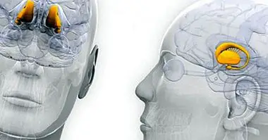
Arachnoid (brain): anatomy, functions and associated disorders
The meninges are a series of membranes that together with the skull and spine protect the central nervous system , so that it prevents minor blows or injuries can alter its operation or destroy it completely.
In addition to this they allow the brain to be in place and, because there are different types of pain receptors in them, we can perceive that there is some type of problem or injury. In this article we are going to explore the arachnoid membrane in order to analyze what it is, its particularities and functions.
- Recommended article: "Parts of the human brain (and its functions)"
Arachnoids: the intermediate meninge
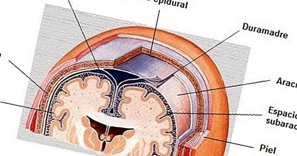
The arachnoid, together with the dura mater and the pia mater, is one of the three meninges . These are membranes that protect our brain and marrow from injuries coming from outside and that have an important role in our survival. They are placed one below the other, forming three small protective layers.
In the case of the arachnoid we are facing the intermediate membrane, being in contact with the dura mater and the pia mater. It turns out to be the most fragile and prone to break of the three. Another of its main characteristics is that it is not vascularized.
Although they are in close contact, the arachnoid is separated from the dura through the subdural space, which is more than a space a thin layer of cells between which interstitial fluid is found. With respect to the pia mater, it is separated from it by the subarachnoid space, and in turn connects with it by means of the arachnoid trabeculae.
One of the main aspects that distinguish it from the other two meninges is the fact that it contains the subarachnoid space, through which the cerebrospinal fluid circulates.
Main components
Observing the arachnoid we can identify the existence in them of different layers or parts.
1. Arachnoid or arachnoidal barrier layer
It corresponds to the part of the arachnoid that is in contact with the dura mater . Their cells are very close and hardly allow the passage of interstitial fluid, being the most resistant part of the arachnoid. This layer prevents ions and molecules from entering or leaving. However, in it, a series of arachnoid granulations or villi can be distinguished by which they connect with the existing veins in the dura, which allows the cerebrospinal fluid to be expelled at the end of its cycle.
2. Arachnoid trabeculae or reticular arachnoid layer
The cells of the arachnoidal barrier layer project towards the pia mater, forming a network that crosses the subarachnoid space which in turn forms a network or mesh that in fact gives name to the meninx (due to the resemblance to the fabric of a spider). Within these projections we find fibers in net, anchor fibers and microfibers. The exact function of the trabeculae is not yet fully known, although it is speculated that they are capable of perceiving the pressure caused by the cerebrospinal fluid.
3. Subarachnoid space
Although more than part of the arachnoid is a space located between its blades, the subarachnoid space is one of the most important parts of the arachnoid. This is so because it is through it that the cerebrospinal fluid passes . In this space we can also find a series of important pits and cerebral cisterns in which the cerebrospinal fluid accumulates and which allow its distribution.
In addition to the brain itself, an orbital subarachnoid space surrounding the optic nerve can be found.
Principal functions
The arachnoid is a membrane that, like the other meninges, has diverse functions that allow and favor our survival.
1. Protects the nervous system
Despite being relatively fragile, the arachnoid, together with the rest of the meninges, allows the brain and spinal cord to be protected against blows and injuries , as well as contamination and infection by harmful agents.
2. Cerebrospinal fluid distribution
It is in the arachnoid and in the different cisterns of the subarachnoid space through which the cerebrospinal fluid circulates , essential element to keep the neurons of the nervous system alive by nourishing them and in turn allowing the elimination of the residues of brain functioning.
3. Vascular-cerebrospinal fluid system connection
Cerebrospinal fluid carries the waste of brain activity, but it is necessary to expel them . This is done through the blood of the veins of the dura mater, with whom the arachnoid communicates. In the same way it prevents the cerebrospinal fluid from accumulating excessively, which does not cease to be secreted.
4. It allows the buoyancy of the brain
The fact that the cerebrospinal fluid circulates through its interior allows the brain to be somewhat floating , which reduces its weight and allows the maintenance of its morphology.
5. Perception of intracranial pressure
Although it is something that is not completely known, it is suspected that they are the arachnoid trabeculae those that allow the body to detect increases in intracranial pressure.
Associated disorders
There are several affectations that are linked to alterations in the arachnoid or in another of the meninges. Among these alterations we can find the following.
1. Arachnoid cyst
Small cystic structures can form within the arachnoid that fill with cerebrospinal fluid. While they may not cause problems, they may generate a pressure that damages the nervous system. Headaches, sensitivity problems, paresthesia or paralysis are common.
2. Meningitis
Both the arachnoid and the rest of the meninges can succumb to a viral or bacterial infection, becoming inflamed and causing different symptoms such as dizziness, headache or weakness. The composition of the cerebrospinal fluid circulating in the arachnoid may be altered , as well as causing compression of the brain.
3. Communicating hydrocephalus
It is a disorder in which cerebrospinal fluid builds up inside the skull , in this case because the parts of the arachnoid that allow the communication between it and the blood of the veins does not work correctly, accumulating too much fluid and not re-entering the blood.
4. Subarachnoid hemorrhage
It occurs when due to illness or injury (such as due to a traumatic brain injury) , the blood enters and floods the subarachnoid space. It can be deadly. Headache, alterations of consciousness and gastrointestinal problems such as nausea and vomiting are frequent.
Bibliographic references:
- Kandel, E.R .; Schwartz, J.H .; Jessell, T.M. (2001). Principles of Neuroscience. Madrid: McGraw Hill.
- Martínez, F .; Tomorrow, G .; Panuncio, A. and Laza, S. (2008). Anatomo-clinical review of the meninges and intracranial spaces with special reference to chronic subdural hematoma. Mexican Journal of Neuroscience; 9 (1): 47-60