Meninges: anatomy, parts and functions in the brain
Regardless of the alarmingly high levels of sedentary lifestyle observed in the population, as a rule human beings are moving continuously.
We walk, we run, we dance, we jump, we interact with the environment and with other individuals ... all these actions can cause that under certain circumstances the organs that are part of our organism, including those of the nervous system, they run the risk of being damaged .
That is why it is necessary the presence of protection systems that keep everything in place and that block the arrival of possible injuries. Fortunately, our body has different structures that allow us to protect our viscera, organs and internal structures. In the case of the nervous system and the brain, it is protected by the skull and spine, along with other structures and elements such as the blood-brain barrier or, in the case at hand, a series of membranes called meninges .
What are the meninges?
Imagine that we are on an operating table and we need to make way for a part of the patient's brain. After crossing a layer of skin and muscle, we would reach the skull, a bone structure that protects the brain. But nevertheless, if we go through this bone protection we do not find ourselves directly with the brain , but we will find a series of membranes that surround the nervous system. These membranes are called meninges.
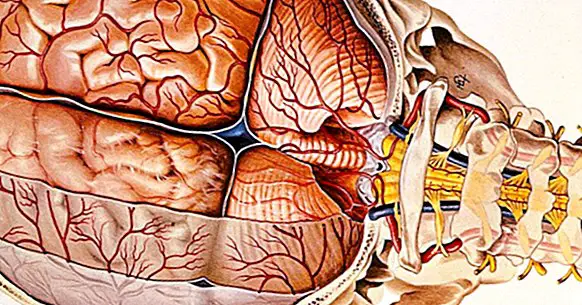
The meninges are a set of protective layers located between the central nervous system and its bone protection , both at the level of the brain and the spinal cord. Specifically, you can find a series of three membranes located one below the other, receiving from more external to more internal the name of dura mater, arachnoid and pia mater . Through them circulate different fluids that contribute to keep clean and nourished the brain, being crossed and irrigated by different blood vessels,
Although when we talk about the meninges, we think fundamentally about the membranes that cover the brain, it is important to point out that these structures they cover the whole central nervous system and not just the brain , also protecting the spinal cord.
The three meninges
As we have indicated previously, we understand as meninges a set of three membranes that internally protect the nervous system.
From more external to more internal, are the following.
1. Dura mater
In addition to being the outermost meninge, the dura is the hardest and most condensed of the three of which we have, and is also the one that is closest to the outside. Attached partly to the skull, this membrane protects the brain and acts as a structural support for the entire nervous system by dividing the cranial cavity into different cells.
In the dura are most of the great blood vessels of the brain , since in addition to protecting them, it allows them to have a space through which to distribute themselves and move from one location to the next. Subsequently, these blood vessels will be diversified into different subdivisions as they deepen in the brain.
- To know more about this layer of the meninges, you can visit this article: "Dura mater (brain): anatomy and functions"
2. Arachnoids
Located in an intermediate area between dura mater and pia mater, the arachnoid is a meninge that receives its name due to its morphological similarity to the fabric of a spider , that is, its grid configuration. It is the most delicate of the three meninges, a transparent and non-vascularized layer attached to the dura mater.
It is mainly because of this meninge and the space between the arachnoid and the pia mater where the cerebrospinal fluid circulates. In addition, it is in the arachnoid that the end of the cerebrospinal fluid life cycle occurs, which is returned to the blood flow through the villi or structures known as arachnoid granulations in contact with the large veins that run through the dura.
3. Piamadre
The innermost, flexible meninges and in greater contact with the structures of the nervous system It's the pia mater. In this layer you can find numerous blood vessels that irrigate the structures of the nervous system.
It is a thin membrane that remains stuck and infiltrates the cerebral folds and convolutions. In the part of the pia mater in contact with the cerebral ventricles we can find the choroid plexuses, structures in which the cerebrospinal fluid that irrigates the nervous system is synthesized and released.
Spaces between the meninges
Although the meninges are located one behind the other, the truth is that some of them can be found among them intermediate spaces through which cerebrospinal fluid flows . There are two intermediate spaces, one between dura mater and arachnoid called subdural space and the other between arachnoid and pia mater, the subarachnoid. It should also be mentioned that in the spinal cord we can find one more space, the epidural space. These spaces are the following.
1. Subdural space
Located between dura mater and arachnoid the subdural space is a very slight separation between these meninges through which interstitial fluid circulates, which bathes and nourishes the cells of the different structures.
2. Subarachnoid space
Below the arachnoid itself and by contacting arachnoid and pia mater we can find the subarachnoid space, through which the cerebrospinal fluid flows. In some areas of the subarachnoid space the separation between arachnoid and pia mater is widened, forming large brain cisterns from which the cerebrospinal fluid is distributed to the rest of the brain.
3. Epidural space
While in the brain the outermost layer of the dura is attached to the skull, inside the spine does not happen the same: in the spinal cord there is a small separation between bone and bone marrow. This separation is what is called epidural space, connective tissue and lipids that protect the marrow are found in it while we move or change position.
It is in this location that epidural anesthesia is injected in women who are in the process of giving birth, blocking the transmission of nerve impulses between the medulla and the lower part of the body.
Functions of the meninges
The existence of the meninges is a great advantage for the human being when it comes to maintaining the functioning of the nervous system. This is because these membranes perform a series of functions that allow adaptation , which can be summarized in the following.
1. They protect the nervous system from physical injuries and other damages
The meningeal system as a whole supposes a barrier and damping element that prevents or hinders that blows, traumatisms or injuries cause serious or irreparable damages to the central nervous system, we are talking about the skull or the spinal cord.
They also act as a filter that prevents harmful chemical agents from entering the nervous system. That is to say, that the meninges offer a protection that consists of a physical and chemical barrier at the same time.
2. It allows the brain to remain healthy and stable
The meninges participate in the genesis and allow the circulation of cerebrospinal fluid, a key element when it comes to eliminating the waste generated by the continuous brain function and maintain intracranial pressure .
Other liquids, such as the interstitial, also circulate through this system, allowing the aqueous medium in which the nervous system is stable. In addition, the blood vessels that supply the brain pass through the meninges, I also feel protected by them. In conclusion, the meninges they act facilitating the survival and nutrition of the nervous system .
3. Keeps the nervous system in place
The presence of the meninges prevents the nervous system from moving too much, fixing the structures that are part of it to a more or less stable situation and causing a fixed internal structure to be maintained , as it happens in the intracranial cavity and its division into cells. This is important, because the consistency of most parts of the nervous system is almost gelatinous and therefore does not have to stay in place.
In short, the meninges act as a belt and give shape and unity to the whole of this part of the nervous system, which allows normal operation.
4. Inform the agency of possible problems
Although the perception of stimuli and internal states of the organism occurs thanks to the action of the nervous system, the central nervous system itself does not have receptors that report internal problems, such as nociceptors. Fortunately, this is not the case with the meninges, which do They have tension, expansion, pressure and pain receptors and by consigiente inform about what happens in that part of the internal environment.
Thus, it is thanks to them that it is possible to capture the existence of neurological problems (regardless of whether these problems cause other perceptual or behavioral problems), being the headaches the product of alterations in these membranes.
Bibliographic references:
- Kandel, E.R .; Schwartz, J.H .; Jessell, T.M. (2001). Principles of Neuroscience. Madrid: McGraw Hill.
- Kumar, V. (2015). Robbins and Cotran Pathologic Mechanisms of Disease. Philadelphia: Elsevier Saunders.
- Martínez, F .; Tomorrow, G .; Panuncio, A. and Laza, S. (2008). Anatomo-clinical review of the meninges and intracranial spaces with special reference to chronic subdural hematoma. Revista Mexicana de Neurociencia: 9 (1): 17-60.
- Tortora, J.G. (2002). Principles of anatomy and physiology. 9th edition. Mexico D.F.; Ed. Oxford, pp. 418-420.

