Optic nerve: parts, path and related diseases
Sight is one of our most essential senses, probably being the most developed exteroceptive sense in the human being. Not surprisingly, we devote a large part of our brain to the processing of visual information, being able to perceive a wide variety of parameters such as color, shape, depth or brightness with a sharpness and precision notorious.
But to be able to process all that information, and in fact to be able to see in general, it is first necessary that the information that the eyes capture reaches the relevant brain nuclei. Y this would not be possible without the existence of the optic nerve , about which we are going to talk next.
- Related article: "The 11 parts of the eye and its functions"
Optic nerve: basic description and location
We give the name optic nerve to a tract or set of nerve fibers that go from the eye to the central nervous system and whose presence allows vision. This tract is part of the cranial nerves, specifically the pair II, and consists of more than a million neurons (approximately an estimated one and a half million) of sensory type, not transmitting information to the eye but only receiving it from him.
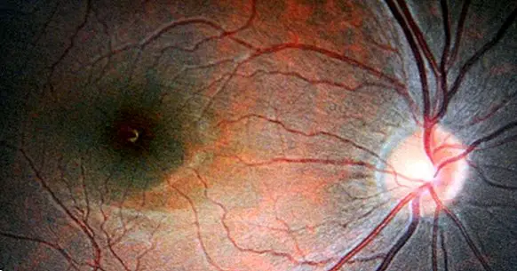
This nerve can be located in a space between the back of the eyeball, taking one of its ends in the ganglion cells of the retina, on the one hand, and the optic chiasm, on the other . This small section, between 4 and 5 cm in length, is of vital importance and without it we could not be able to see.
From the chiasm most of the fibers of the optic nerves of both eyes will decuss (that is, the left eye will go to the right hemisphere and vice versa), forming a tract that will go to the lateral geniculate nucleus and from there to different nuclei of the cerebral cortex.
The optic nerve has the peculiarity that initially the fibers that will shape it (the neurons that connect with the ganglion cells) are not myelinated until they meet in the so-called optic disc or blind spot, an area where there are neither cones nor rods and from which the neurons will form the optic nerve itself, already myelinated in order to allow a rapid and efficient transmission of visual information.
So, the optic nerve, which It is mainly formed by myelinated axons , it is mainly white matter. Although it originates outside the skull (in the retina), once entered into this and especially the bony part, the optic nerve is covered and protected by the meninges.
- Maybe you're interested: "Cranial nerves: the 12 nerves that come out of the brain"
What is it for?
The main function of the optic nerve, as you can guess, is to transmit the visual information that we capture through the photoreceptors of the retina to the rest of the brain in order to process and interpret it.
First the photoreceptor captures the outside information , generating a series of electrochemical reactions that in turn will transform the data into bioelectric impulses that will activate the ganglion cells of the retina, which in turn will travel to the blind spot where the nerve fibers join to form the optic nerve, the which will proceed to send the message.
Interestingly, despite being perhaps the nerve that has greater importance when it comes to seeing its location in the retina is what causes the existence of our blind spot.
Parts of the optic nerve
Although the optic nerve is relatively small in its journey to the optic chiasm, the truth is that you can observe different segments in your journey between the eye and the aforementioned chiasma . Among them, the following stand out.
1. Intraocular segment
This first segment of the optic nerve is the one that still occurs inside the eye, in the section that It goes from the ganglion cells to the blind point and then passes through the lamina or cribriform area , which crosses the sclera and the choroid.
2. Intraorbital segment
It is the part of the optic nerve that goes from the exit of the eye to its exit from the eye orbits. In this part the nerve it goes around the muscles that control the eye and the fat after her.
3. Intra-acicular segment
In this third segment it is in which the optic nerve reaches the skull at the end, next to the ophthalmic artery. For this the nerve enter through a hole called optic foramen . This area is one of the most sensitive and easy to injure.
4. Intracranial segment
The last segment is the intracranial, in which the optic nerve is already completely inside the skull and travel to the optic chiasm. This is where he receives the protection of the meninges.
Pathologies and problems associated with your injury
The optic nerve is one of the most important of our visual and is that without it, vision as such would not be possible. There are many possible conditions that can occur in this nerve and cause us either blindness or alterations and difficulties in the vision.
Among them we can find the atrophy of the optic nerve derived from for example a neuropathy (for example derived from metabolic problems such as diabetes), intoxication, meningitis (remember that the meninges cover this nerve in some portions, so in case of inflammation they could compress and damage it), strokes or tumors that generate pressure or destroy that nerve.
Another possibility is that the nerve itself is inflamed, a condition called optic neuritis that is usually linked to infections and autoimmune problems. There may also be accumulations of substances that form the so-called harsh, especially in the head of the optic nerve (the area in which it starts in the blind spot).
Finally, and probably the most well-known and frequent problem that can generate blindness linked to the optic nerve, is glaucoma . This disease is derived from a progressive increase in intraocular pressure, which progressively damages the nerve.
Bibliographic references:
- Miller, N.R. & Newman. N.J. (eds) (2005) .. Walsh and Hoyt's clinical neuro-opthalmology. 6th edition. Baltimore: Williams & Wilkins, 385-430.
- Sánchez, F. (2001). The optic nerve and vision disorders. Integral Medicine, 38 (9): 377-412. Elsevier